Background
Peritoneal mesothelioma is a rare neoplastic disease affecting the mesothelium. It could originate in the mesothelium of the pleural, peritoneal, and pericardial cavities, and in the testicular tunica vaginalis. Multicystic peritoneal mesothelioma (MCPM) represents 3%-5% of all peritoneal mesotheliomas. The estimated incidence is about 2 in 1,000,000 per year [1,2]. The pathophysiology is unknown, although, it occurs predominantly in young women, with a previous history of endometriosis. MPCM involves different histologic types: low-grade, well-differentiated papillary, multicystic, epithelioid, sarcomatoid, biphasic (epithelioid and sarcomatoid), and deciduoid [3]. Subacute abdominal discomfort is the common clinical manifestation of MCPM, however, it is typically diagnosed as an incidental finding. The treatment initially consists of the complete surgical excision of the abdominal masses [4]. Short-term survival appears to be favorable, although no long-term clinical trials have been reported. Along with complete surgical removal of the masses, hyperthermic intraperitoneal chemotherapy (HIPEC) is reported to be effective for diffuse malignant peritoneal mesothelioma [5-7]. We report a case of MCPM presented with acute abdominal pain.
Case Presentation
We describe a case of a 48-year-old man presenting with persistent severe abdominal pain at the Emergency Department of the Ospedale Castelli of Verbania. He reported no relevant past clinical history. The physical examination of the abdomen revealed diffuse abdominal pain, particularly in hypogastrium. First, he underwent blood tests and abdominal ultrasound which were inconclusive. We performed a complete abdominal computed tomography (CT) scan which described peritoneal effusion predominantly mesogastric and hypogastric and in the pelvis, in contiguity with oval pluriconcameral septate formation with a maximum diameter of about mm 75 regular margins, hyperdense with suprahydric density content. The formation appeared cleaved by the seminal vesicles, apparently in close connection with the left lateral wall of the sigmoid colon with a cleavage plane that is not well recognized (Figures 1 and 2).
After the first diagnosis approach, our differential diagnosis were mesenteric cyst; pseudomyxoma peritonei, cystic teratoma, and peritoneal mesothelioma. We considered the clinical examination and the radiological imaging, and we decided that a diagnostic laparoscopy to be the best approach. The procedure was performed with a 12-mmHg pneumoperitoneum through a Veress needle placed in the left hypochondrium. We placed a 12-mm disposable optic trocar in the right paraumbilical site with an advancement of a 10 mm 30-degree angled laparoscope. On preliminary exploration of the abdominal cavity, we detected a large amount of free citrine effusion and numerous cystic formations located predominantly in the pelvis and in the right parieto-colic gutter. No evidence of Glissonian outcropping liver lesions or other nodular lesions of the abdominal wall or bowel was detected. Placement under the vision of two additional disposable trocars: 12 mm in the right flank and 5 mm in the left flank. We proceeded with the aspiration of the free effusion and sent a sample to Pathology Anatomy for the cytological examination. In the pelvis, we found two cysts tenaciously adhered to the parietal peritoneum and sigmoid colon and rectum. We performed careful dissection of the cysts from the bowel wall and the parietal peritoneum (Figure 3).
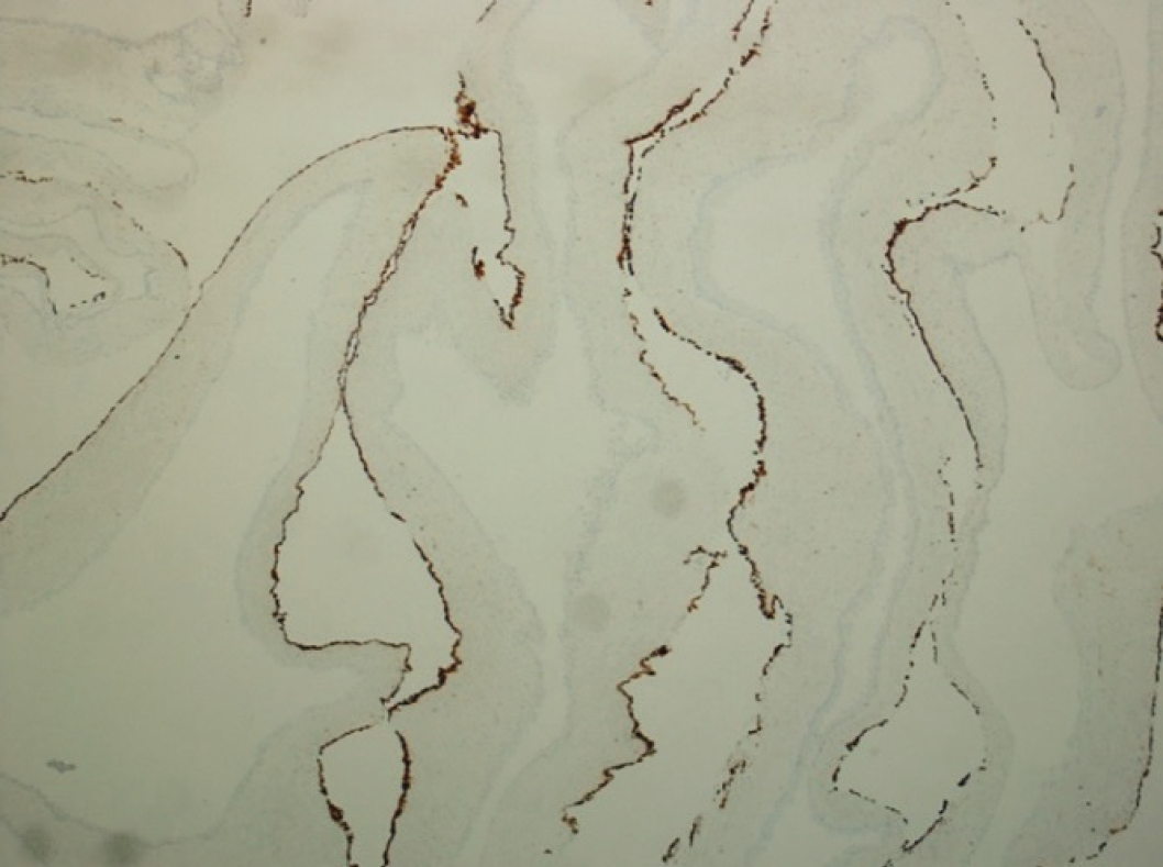
We subsequently inserted the cysts inside of an endobag and extracted them through the paraumbilical site. We sent multiple cysts for definitive histology. The diagnosis turned out as MCPM. Microscopically, the cysts showed a flattened - cuboidal or micropapillary mesothelial lining with an extensive amount of squamous metaplasia; focally, cyst walls also showed fibrosis and an intense inflammatory infiltrate, with hemorrhagic areas. The immunohistochemical exam reported positivity to Calretinin, CK5/6, CK7, Ki67, p40 in areas with squamous metaplasia, p63, and WT1 (Figures 4 and 5).
The postoperative course was free of complications, the patient was discharged after 5 days. He was extensively informed about the need to pursue follow-up at a specialized center. A control CT scan was performed after 6 months, and it revealed no signs of recurrence. The patient is still on follow-up, but he refused every other invasive treatment, like HIPEC.
Discussion
The natural history of this rare disease is still a matter of debate. It is considered to be an intermediate grade between benign and malignant peritoneal mesothelioma [8,9]. Despite the short-term survival rate being favorable, the recurrence rate is high, even in patients who underwent complete surgical excision [10]. The recurrence rate after 2 years is estimated to be approximately 50% [11]. The malignant transformation of benign cystic peritoneal mesothelioma is also described, even after complete surgical excision [12]. Currently, there is no universal consensus regarding the surgical approach. Some authors prefer conservative treatments such as irradiation, percutaneous cyst drainage, hormone-therapy, and sclero-therapy with anthracycline and finally, only radiological follow up [8]. Although such treatments are described in the literature, the safest and most effective treatment seems to be the association of cytoreductive surgery and HIPEC. It is also the treatment of choice in case of recurrence. The rationale behind the use of HIPEC is the eradication of microscopic residual tumors which could decrease the risk of recurrence [11].

Calretinin immunoreaction shows strong staining of the mesothelial cells (CONFIRM anti-Calretinin clone SP65, Ventana).
We performed a complete excision of the masses during the intervention as recommended. The CT scan revealed no radiological signs of recurrence. Although current guidelines advise for cytoreductive surgery such as HIPEC, the patient refused every other invasive treatment. The patient is still on follow-up at a specialized clinical hospital, no clinical and radiological signs of recurrence have been diagnosed after 18 months.
Conclusion
MPCM is a neoplasm whose pathophysiology is yet unknown, whose behavior is barely understood, and for which there isn’t yet a general agreement on its management. This is due to the lack of longitudinal studies and its extremely low rate of incidence. Our experience demonstrates no signs of recurrence after one year from the intervention, without more invasive surgical treatment. This raises the issue of the clinical utility of undergoing further invasive procedures for this rare disease. Long-term clinical trials are necessary to further improve the management of the MCPM.


