64
views
0
recommends
+1 Recommend
1 collections
    0
    shares
      scite_
      0
      0
      0
      0
      Smart Citations
      0
      0
      0
      0
      Citing PublicationsSupportingMentioningContrasting
      View Citations

      See how this article has been cited at scite.ai

      scite shows how a scientific paper has been cited by providing the context of the citation, a classification describing whether it supports, mentions, or contrasts the cited claim, and a label indicating in which section the citation was made.

       
      • Record: found
      • Abstract: found
      • Article: found
      Is Open Access

      Prevention of recurrence in malignant proliferating trichilemmal tumor of scalp in young female using adjuvant radiation therapy after surgery

      Published
      case-report
      Bookmark

            Abstract

            Background:

            Proliferating trichilemmal tumors (PTTs) are rare benign neoplasms that arise from the outer sheath of a hair follicle. Group III PTTs are high-grade malignant tumors reported to exhibit a high recurrence rate, lymph nodes, and a tendency to develop distant metastasis.

            Case Presentation:

            A 25 years old female patient was presented to us with a history of recurring soft tissue scalp lesions. The lesion recurred three times within 1 year. There was no history of trauma. Every time the lesion was excised but no histopathology of the specimen was done. Upon the third recurrence, it came out to be a malignant proliferating trichilemmal tumor (MPTT). Considering the young age of the patient and keeping in view the history of multiple recurrences with positive deep margin even after the third surgery, the patient was planned external beam radiotherapy (RT) 50 Gy/25 fx to scalp with 9 Mev electron using 0.5 cm bolus. The post-RT follow-up currently of 2 years is uneventful.

            Conclusion:

            The mainstay of treatment is surgery with wide surgical margins as local recurrence is most common in MPTT. Adjuvant RT was used in this case to prevent recurrence.

            Main article text

            Background

            Proliferating trichilemmal tumors (PTTs) are rare benign neoplasms that arise from the outer sheath of a hair follicle [1]. Only a few hundred cases have been described in the literature [2]. Accordingly, Ye et al. [3] classified PTTs into three groups, namely, benign, low-grade malignant, and high-grade malignant lesions, based on clinic-pathological characteristics. Group I PTTs are benign lesions. Recurrence is not observed in these lesions. They show histologically regular contours with surrounding tissues and mild nuclear atypia but do not involve increased mitotic activity, necrosis, and lymphovascular invasion. Group II PTTs are low-grade malignant tumors. Local recurrence may be observed in these lesions. These tumors have histologically irregular and local invasive contours, and they elongate to the deep dermis and subcutaneous tissue. Group III PTTs are high-grade malignant tumors reported to exhibit a high recurrence rate, lymph nodes, and a tendency to develop distant metastasis. Remarkable nuclear pleomorphism, atypical mitosis, necrosis, and lymphovascular invasion may be observed in these tumors.

            Case Report

            A 25-year-old female patient was presented to us with a history of recurring soft tissue scalp lesions. The lesion recurred three times within 1 year. There was no history of trauma. The patient got an excision of a 2 × 2 cm scalp lesion twice and histopathology of the specimen was not done. On the third recurrence, lesion was excised and diagnosed as a malignant proliferating trichilemmal tumor (MPTT) histopathologically. She had a scalp lesion of T size; 1.9 × 1.6 × 1.5 cm. The tumor was 0.7 cm from the first polar resection margin, 0.6 cm from the second polar resection margin, 0.1 cm from deep margins, and less than 0.1 cm from the peripheral resection margins. It was Grade III, lymphovascular invasion and perineural invasion was not identified. Nodal and distant metastatic status was unknown. She was referred for redo surgery to achieve margins.

            When she came back after surgery, her histopathology report showed residual MPTT of T size: 0.8 × 0.6 × 0.5 cm, and mitosis was approximately 15/10 under high power field. All margins were free of tumor involvement on the frozen section, but the tumor was 0.1 cm away from the closest deep margin. Additional pathological findings included foci of chronically inflamed granulation tissue and foreign body-type multinucleated giant cell reaction was seen. These findings were consistent with previous surgical sites.
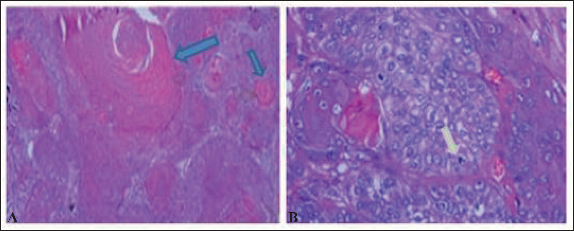
            Figure 1.

            Histopathology of MPTT of scalp showing. (a) Trichilemmal keratinization represented by blue arrow (b) Tumor cell with wide cytoplasm represented by the light grey arrow.
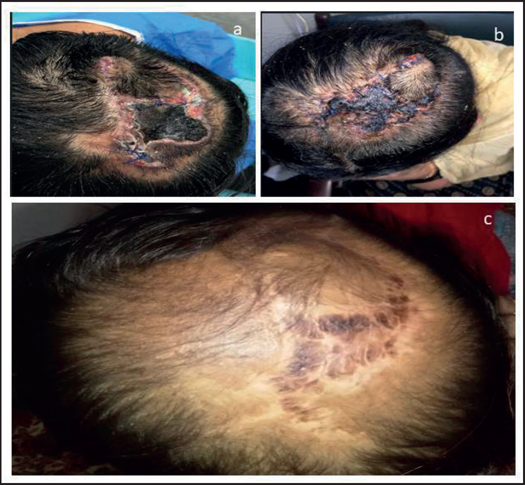
            Figure 2.

            (a) After surgery. (b) 2 weeks after radiation therapy. (c) Patient on 9 months follow up with healed scalp and no recurrence.

            Considering the young age of the patient and keeping in view the history of multiple recurrences and deep margin of 0.1 cm, the patient was planned scalp radiotherapy (RT) 50 gy/25 fx with 9 Mev electron using 0.5 cm bolus. The patient completed RT, and now she is on routine follow-up (Figure 2).

            Discussion

            MPTT is a rare malignancy, usually observed in the sixth decade of life [4]. The tumor may be misdiagnosed as squamous cell carcinoma (SCC) because cutaneous SCC is commonly observed in the scalp [5]. Moreover, the histological features of MPTT and SCC are more or less similar. It is commonly characterized by abnormal mitosis and infiltrating margins [5].

            Similarly, benign and malignant PTT may also be confused and is important to differentiate. Three major groups have been identified in PTT namely benign, low-grade malignant, and high-grade malignant tumors [6]. Benign lesions usually do not have a recurrence while low- and high-grade lesions have low and high recurrence rates respectively. MPTT is generally transformed from a benign PTT but may appear as a malignant tumor de novo in a few cases [3].

            According to the literature, >80% of cases of MPTT are observed in elderly women [7]. Our patient is a young female only 25 years of age. She is having high-grade MPTT and neoplasm has recurred thrice and the tumor has a high mitotic activity (Figure 1). All these features reflect high-grade malignant disease.

            A mainstay of treatment is surgery with wide surgical margins as local recurrence is most common in MPTT [8]. Patients are usually closely monitored after surgery for any possible relapse of disease. Adjuvant RT was used in this case to prevent recurrence. Our patient is disease free on routine follow-up for 1 year after adjuvant RT.

            What is new?

            PTTs are rare. Only a few hundred cases have been described in the literature. The mainstay of treatment is surgery with wide surgical margins as local recurrence is most common in MPTT. In our study, considering the young age of the patient and keeping in view the multiple recurrences and deep margin of 0.1 cm, the patient was planned scalp RT 50 gy/25 fx with 9 Mev electron using 0.5 cm bolus. Our patient is disease free on routine follow-up for 1 year after adjuvant RT.

            List of Abbreviations

            fx

            Fraction

            gy

            Gray

            MPTT

            Malignant proliferating trichilemmal tumor

            PTTs

            Proliferating trichilemmal tumors

            SCC

            Squamous cell carcinoma

            RT

            Radiotherapy

            Conflicts of interest

            The authors declare that they have no conflict of interest regarding the publication of this case report.

            Funding

            None.

            Consent for publication

            Written informed consent was obtained from the patient.

            Ethical approval

            Ethical approval was taken from the Institutional Review Board of INMOL Hospital Lahore (Reference no.: INM/PA/2073, dated: August 6, 2021). Prior patient consent was taken for the publication of the study.

            References

            1. Alici O, Keles MK, Kurt A. A rare cutaneous adnexal tumor: malignant proliferating trichilemmal tumor. Case Rep Med. 2015. 1–4. [Cross Ref]

            2. Chaichamnan K, Satayasoontorn K, Puttanupaab S, Attainsee A. Malignant proliferating trichilemmal tumors with CD34 expression. J Med Assoc Thai. 2010. Vol. 93(Suppl 6):S28–34

            3. Ye J, Nappi O, Swanson PE, Patterson JW, Wick MR. Proliferating pilar tumors: a clinicopathologic study of 76 cases with a proposal for definition of benign and malignant variants. Am J Clin Pathol. 2004. Vol. 122(4):566–74. [Cross Ref]

            4. Gallant JN, Sewell A, Almodovar K, Wang Q, Dahlman KB, Abramson RG, et al.. Genomic landscape of a metastatic malignant proliferating tricholemmal tumor and its response to PI3K inhibition. NPJ Precis Oncol. 2019. Vol. 3:5[Cross Ref]

            5. Parambeth HK, Udhayam N, Agarwal S, Gupta S, Giridhar P, Rath GK. A large helmet-shaped proliferating trichilemmal tumor of the scalp: is definitive radiotherapy the treatment? Acase report. J Egypt Nat Cancer Inst. 2019. Vol. 31:7[Cross Ref]

            6. Sau P, Graham JH, Helwig EB. Proliferating epithelial cysts. Clinicopathological analysis of 96 cases. J Cutan Pathol. 1995. Vol. 22(5):394–406. [Cross Ref]

            7. Deshmukh BD, Kulkarni MP, Momin YA, Sulhyan KR. Malignant proliferating trichilemmal tumor: a case report and review of literature. J Can Res Ther. 2014. Vol. 10:767–9. [Cross Ref]

            8. Dubhashi SP, Jadhav SK, Parasnis A, Patil CS. Recurrent malignant proliferating trichilemmal tumor with lymph node metastasis in a young woman. J Postgrad Med. 2014. Vol. 60(4):400–2. [Cross Ref]

            Summary of the case

            1 Patient (gender, age) 25 years, female
            2 Final diagnosis Malignant proliferating trichilemmal tumor of scalp
            3 Symptoms Scalp lesions
            4 Medications Symptomatic treatment given
            5 Clinical procedure Surgery followed by radiotherapy
            6 Specialty Oncology

            Author and article information

            Journal
            European Journal of Medical Case Reports
            EJMCR
            Discover STM Publishing Ltd.
            2520-4998
            30 June 2023
            : 7
            : 6
            : 118-120
            Affiliations
            [1 ]Department of Radiation Oncology, INMOL Cancer Hospital, Lahore, Pakistan
            Author notes
            [* ] Correspondence to: Sana Naeem Department of Radiation Oncology, INMOL Cancer Hospital, Lahore, Pakistan. syedasananaeem@ 123456gmail.com
            Author information
            https://orcid.org/0000-0002-9116-0899
            Article
            ejmcr-7-118
            10.24911/ejmcr/173-1669004762
            48e1535c-48cc-4055-93f7-0c6cab0f2814
            © Sana Naeem, Iqra Iftikhar, Rab Nawaz Maken, Misbah Masood, Abubaker Shahid

            This is an open access article distributed in accordance with the Creative Commons Attribution (CC BY 4.0) license: https://creativecommons.org/licenses/by/4.0/) which permits any use, Share — copy and redistribute the material in any medium or format, Adapt — remix, transform, and build upon the material for any purpose, as long as the authors and the original source are properly cited.

            History
            : 21 November 2022
            : 16 June 2023
            Categories
            CASE REPORT

            Proliferating trichilemmal tumors,scalp lesion

            Comments

            Comment on this article