Background
Invasive ductal carcinoma (IDC) is a diverse breast disease having different subtypes and accounts for 55% of all invasive breast cancers in women [1]. IDC is usually detected on mammography and/or ultrasonography (USG) and confirmed by histopathological examination. However, in resource-constrained settings where access to standard imaging modalities is limited and awareness is low, a large number of breast cancer cases are detected only in advanced stages [2]. Given the increasing burden of breast cancer, efforts aimed at simpler and more effective primary detection methods are encouraged to detect breast malignancies early; thus, prompting early treatment, lowering treatment costs, and improving health status post-treatment.
Clinical trials in minimally resourced settings have validated the suitability of Thermalytix – an AI-based screening solution in detecting breast cancer in symptomatic and asymptomatic women [3]. Here, we present two cases of IDC, both of which were detected by Thermalytix and validated by the pathological investigation.
Case Presentation
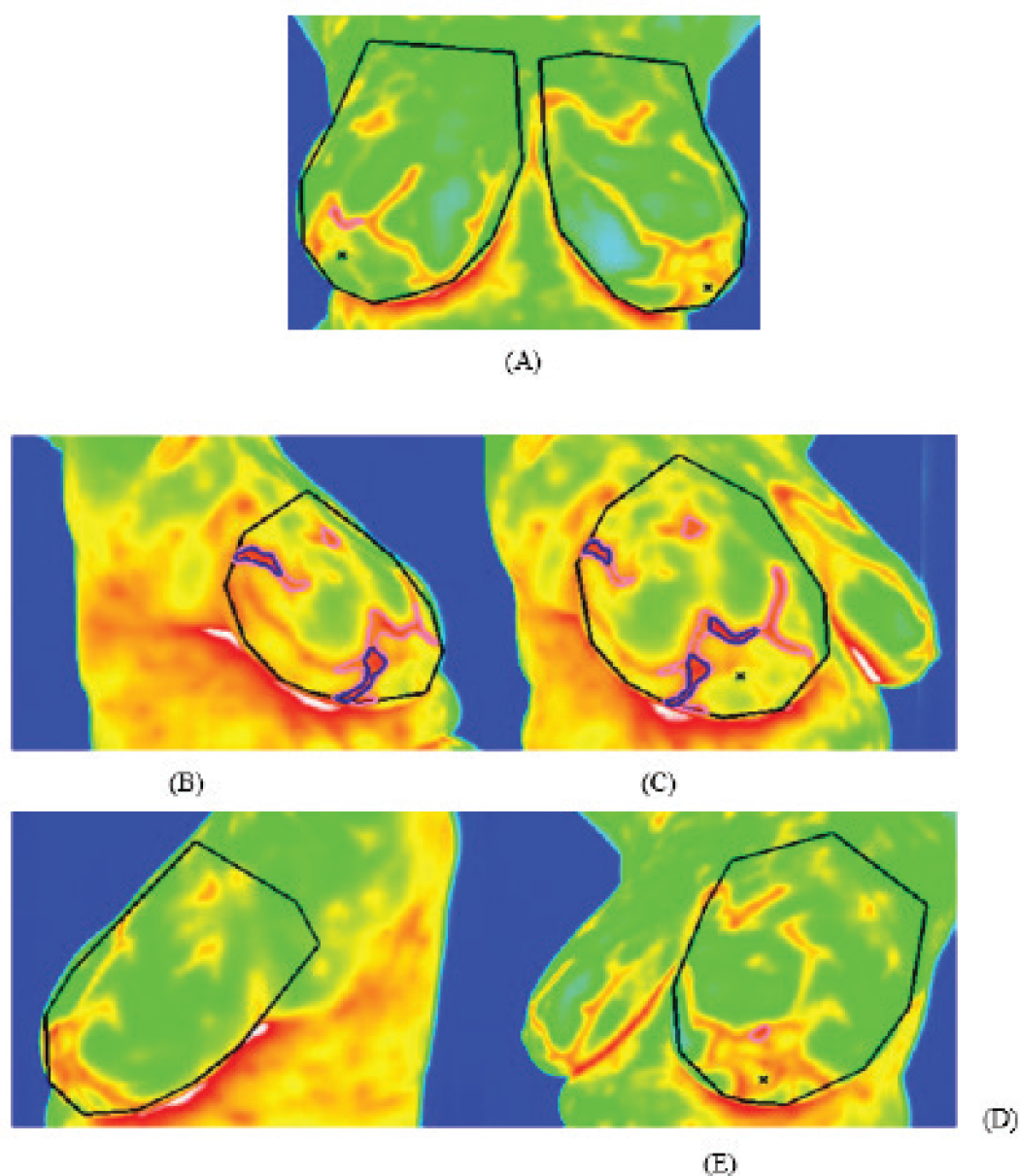
Case 1 : A 47-year-old post-menopausal woman presented with a 3-month history of a lump in the right breast at the 12 O’clock position. Her medical history was unremarkable, and she had no first-degree relatives with cancer. Thermalytix test, an artificial intelligence (AI)-based computer-assisted diagnostic tool, was used to assess the patient. Following the Thermalytix test, a clinical breast examination was performed, and the presence of a palpable lump in the right breast at the 12 O’clock position was noted. During the Thermalytix test, five breast thermal images were captured and analyzed by the automated AI tool. The detailed procedure for the development of the algorithm and scoring is described elsewhere [4]. The test revealed three hotspots, defined as areas of elevated temperature relative to the temperature associated with surrounding tissue, covering 2.3% of the region of interest. In comparison to the surrounding region, the hot spot had a temperature increase of 1.32°C and was irregular and distorted. This hotspot coincides with the lump location creating an impression of a hot lump in the right breast There was no symmetry in breast temperature distribution (Thermal symmetry: 0%), however, the areolar complex showed 100% symmetry (Figure 1). The test also detected six major vessels with five vessel branches on the right breast.

Thermalytix system generated images for case ID B146 showing malignant breast lesions. Images were captured from five different angles: frontal (A), lateral (Right-B, Left-D), and oblique (Right-C, Left- E) views. The regions with pink borders denote warm spots whereas blue borders denote suspicious malignant lesions.
Using the hotspot, areolar, vascular, and overall thermal and/or demographic properties, Thermalytix generated thermobiological, areolar, vascular, and ensemble scores of 0.32, 0.02, 0.69, and 0.66, respectively. A score >0.5 indicated a high likelihood of abnormal lesions. These scores are further combined to obtain a B-Score of 4 indicating a high likelihood of a cancerous lesion in the right breast and a breast ultrasound was recommended for further diagnosis. Table 1 indicates the significance of the B Score and a comparison with the American College of Radiology-Breast Imaging Reporting and Data Systems (ACR BI-RADS) assessment category.
The patient underwent a bilateral mammogram and USG of the right breast lesion. Bilateral mammography evaluation categorized breast density as BI-RADS A parenchymal pattern and detected a well-defined lesion measuring “20 × 20 mm” in the upper outer quadrant of the right breast at 10 O’clock position with mildly spiculated margins and with few microcalcifications. The right axillary nodes were enlarged, with the largest node measuring “18 × 12 mm”. The final impression of the lesion was categorized as ACR BI-RADS 4. USG of the right breast revealed spiculated hypoechoic mass lesion measuring “16 × 12 mm” in the 12 O’clock position Circle II Zone B of malignant etiology and was categorized as BI-RADS 4C lesion - moderate to a substantial likelihood of malignancy 51%-95%.
The histopathology of a wide local excision specimen of the right breast mass showed invasive carcinoma - no special type with lymphovascular invasion. The tumor was grade 2, measuring “20 × 18 × 18 mm” and the resection margins were clear. Dissection of the axillary lymph nodes showed micrometastasis in one of five lymph nodes. The pathological staging for this patient was pT1cN1mi. Immunohistochemical evaluation revealed estrogen receptor positivity, progesterone receptor positivity, HER 2 negativity, and Ki-67 proliferative index up to 25%.
Case 2 : A 46-year-old premenopausal woman presented to a tertiary care hospital with a lump in the upper inner quadrant (UIQ) of her right breast for 2 months. She was the mother of four breastfed children and had no family history of cancer.
The patient underwent the Thermalytix test. Three hotspots were identified that extended to 45.9% of the UIQ of the right breast. The hotspots were irregular and distorted with a 1.14°C increase with respect to the surrounding region. There was neither thermal nor areolar symmetry (0%) and six major vessels with five vessel branches are seen on the right breast.
| B-SCORE | THERMALYTIX INTERPRETATION | ACR BI-RADS ASSESSMENT CLASSIFICATION |
|---|---|---|
| B-Score 0 | Inconclusive. Repeat Thermalytix test, probably an error in thermal image capture. | Category 0: Incomplete – need additional imaging evaluation and/or prior mammograms for comparison |
| B-Score 1 | Normal. No evident signs of breast malignancy in the thermal image. | Category 1: Negative |
| B-Score 2 | Normal. No evident signs of breast malignancy in the thermal image. However, some regions of thermal activity are observed. | Category 2: Benign |
| B-Score 3 | Borderline chances of malignancy. Suggest repeating the Thermalytix test in 3-6 months. For patients with a family cancer history and other risk factors, suggest a diagnostic workup. | Category 3: Probably benign |
| B-Score 4 | Suspicious for malignancy, suggest diagnostic workup | Category 4: Suspicious |
| B-Score 5 | Highly suspicious for malignancy, suggest diagnostic workup | Category 5: Highly suggestive of malignancy |

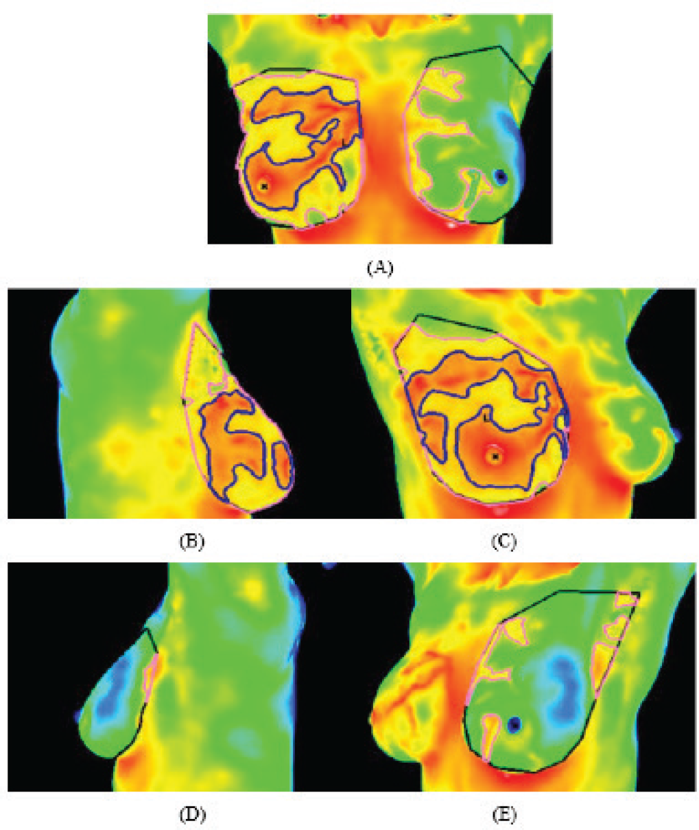
Thermalytix system generated images for case ID: M33 showing malignant breast lesions. Images were captured from five different angles: frontal (A), lateral (Right-B, Left-D), and oblique (Right-C, Left-E) views. The regions outlined with pink borders denote warm spots whereas regions outlined with blue borders denote suspicious malignant lesions.
The high Thermalytix risk scores (TRSs; thermobiological: 0.85, areolar: 0.95, vascular: 0.63, and ensemble: 0.91) and a high B-score of five indicated a high suspicion of a cancerous lesion in the right breast. The AI tool also generated annotated thermal images that showed multiple focal thermal patterns with a thermal increase near the areolar region in the right breast (Figure 2).
Bilateral mammographic evaluation revealed that both breasts were composed of mixed fat and moderate fibro glandular parenchyma. The breast density was categorized as BI-RADS type C. There was no evidence of focal lesion, micro- or macro-calcification, skin thickening, or nipple retraction in both right and left breasts. Mammographic impression was provided as ACR BI-RADS 0, recommending further investigation. An ultrasound guided fine needle aspiration cytology of the right breast lump revealed paucicellular smears. A core biopsy of the right breast was performed to further investigate the lump. The pathology examination results reported the lesion as grade 2 IDC without angioinvasion.
Thus, this was a case of malignancy that was accurately identified by Thermalytix although it was BI-RADS 0 on mammography.
These are unique cases where a malignant breast lesion was detected by Thermalytix and was confirmed on histopathological examination. Table 2 summarizes the characteristics of two cases and Table 3 summarizes the thermal characteristics of the hot lump presented in this report. Thus, a comprehensive Thermalytix-based approach has the potential to identify malignant breast lesions such as IDC.
Discussion
Mammography is the WHO-recommended modality for screening in developed countries [5]. However, mammography may not be the most appropriate screening modality in low- and middle-income countries (LMIC). Mammography also has low sensitivity in women with dense breasts and is usually complemented by USG or magnetic resonance imaging to increase the early identification of malignancies in dense breasts [6]. As these tests are resource-intensive and expensive, the benefits and risks of screening mammography have been debated in LMICs.
Thermography is a medical imaging technique that records changes in the surface temperature of a human body using infrared radiation generated by the body’s surface [7]. Currently available diagnostic modalities (mammography and USG) only detect morphologic changes in the breast once it has grown to a particular size, whereas thermography identifies physiological changes in the breast tissue, such as temperature and blood flow, that are indicative of tumor growth [7]. Thermography, a non-invasive method of breast imaging, was approved as an adjunct imaging modality to mammography in 1982 by The US Food and Drug Administration. However, thermography was not widely accepted due to the complexity involved in interpreting thermograms with the naked eye and resulting in a high false-positivity rate [8]. In recent years, infrared breast thermography is re-emerging as a modality with promising results for detecting breast cancer because of better thermal cameras, improved algorithms, and reduced manual error by using AI [9].
| CASE ID | AGE | SYMPTOMS | MENO-PAUSE | BREAST DENSITY | THERMALYTIX | MAMMOGRAPHY | HISTOPATHOLOGY |
|---|---|---|---|---|---|---|---|
| B146 | 47 | Lump | Yes | A | B-Score 4positive | BI-RADS 4 (Suspicious of malignancy) | IDC Grade 2 |
| M33 | 46 | Lump | No | C | B-Score 5 positive | BI-RADS 0 (Inconclusive) | IDC Grade 2 |
| THERMALYTIX SCORES | B146 | M33 |
|---|---|---|
| Thermo biological scorea | 0.32 | 0.85 |
| Areolar scorea | 0.02 | 0.95 |
| Vascular scorea | 0.69 | 0.63 |
| Ensemble scorea | 0.66 | 0.91 |
| Temperature | 1.32°C increase wrt surrounding region- hot lump | 1.14°C increase wrt surrounding region- hot lump |
score >0.5 indicates a high likelihood of the presence of abnormal lesions.
| MEDICAL SOFTWARE DEVICE | PERFORMANCE | REFERENCE |
|---|---|---|
| Thermalytix Niramai health analytix | Sensitivity 91.02% Specificity 82.39% | Kakileti et al. [10] |
| Transpara™(ScreenPoint Medical/Incepto™) | Sensitivity 86% Specificity 79% | Rodríguez-Ruiz et al. [11] |
| Mammography intelligent assessment® | Sensitivity 85%-97% Specificity 50%-94% | Sharma et al. [12]. |
| Therapixel Nice, France | Sensitivity 69% Specificity 73% | Pacilè et al. [13] |
In our technique, 31 features are extracted from the hot spot region of interest. And to differentiate the malignant heat patterns, we use a random forest (RF) classifier with 200 decision trees. To assess the vascular score, we extract 21 features from vessels detected in both the breast regions. These features are then fed to an RF classifier with 200 decision trees to calculate the Ensemble score. To obtain the final TRS, we combine the proposed vascular score, and hotspot score with critical symptoms like lump and nipple discharge each with different weightages which is explained in the study of Kakileti et al. [4].
Table 4 depicts the comparison of different AI-based screening solutions in terms of their performance. In recent times, thermography has been recommended as a low-cost imaging modality and an alternative to mammography for the early detection of breast cancer lesions in economically backward rural settings [14].
Thermalytix, an AI-based thermography screening tool, was compared to standard of care diagnostics in a multi-site clinical trial of women with symptomatic breast lesions and found to be non-inferior to mammography. Multiple studies have shown the overall sensitivity and specificity of Thermalytix are high [3,10]. The above-described cases depict the technical advances in thermography, coupled with machine learning to improve its diagnostic ability and use as a potential adjunctive tool along with standard imaging modalities.
Conclusion
IDC is the most frequent type of breast cancer and has a high survival rate when diagnosed and treated early. Thermalytix is a portable, radiation-free breast cancer screening technology that uses AI to analyze thermal images. Clinical trials have shown that it is non-inferior to mammography and is a potential screening solution in LMICs. Thermalytix is an automated, affordable, privacy-aware solution for women of all age groups. The analysis of the above two clinical cases of IDC suggests that Thermalytix has the potential in detecting invasive breast cancer in women undergoing breast screening.
What is new?
The presented work evaluates a new technique for the early detection of breast cancer, called Thermalytix, which is a computer-aided diagnostic solution that uses artificial intelligence algorithms over thermal images to detect possible malignant lesions. In this work, the authors describe two clinical cases of symptomatic female patients with invasive ductal carcinoma accurately detected by Thermalytix and further evaluated and confirmed by standard of care imaging and tissue diagnosis methods. The authors believe that this case report will be of interest to your readers because Thermalytix is a novel screening tool that is low-cost, accurate, automated, and portable, allowing it to be used in any clinic.

