82
views
0
recommends
+1 Recommend
1 collections
    0
    shares
      scite_
      0
      0
      0
      0
      Smart Citations
      0
      0
      0
      0
      Citing PublicationsSupportingMentioningContrasting
      View Citations

      See how this article has been cited at scite.ai

      scite shows how a scientific paper has been cited by providing the context of the citation, a classification describing whether it supports, mentions, or contrasts the cited claim, and a label indicating in which section the citation was made.

       
      • Record: found
      • Abstract: found
      • Article: found
      Is Open Access

      Bregmatic sutural bone: a case report

      Published
      case-report
      Bookmark

            Abstract

            Background:

            Sutural bones or wormian bones (an unusual anatomical variant associated with inadequate closure of suture) are extra intrasutural irregular isolated bones that occur additionally at the center of ossification of the skull; along sutures, around sutures or between the cranial sutures formed by bones of the skull vault.

            Case Presentation:

            This report displays a case of the presence of an unusual sutural bone at the bregma of the skull of a cadaver during maceration; hence revealing and supporting the possibility of variations in ossification centers and skull matrix which could aid neurosurgeons, orthopedics, and radiologists in their practices.

            Conclusion:

            The human bregmatic sutural bone presented in this report is a rare occurrence. Considering its location in the skull, it is the first kind reported in Nigeria and Africa as well as one of the few cases reported across the Globe.

            Main article text

            Background

            Bregmatic sutural bone is an anatomical deviant of the skull that is associated with deficient closure of the suture at the bregma [1]. Despite the unusualness of sutural bone also known as wormian bone, intrasutural bone, or supernumerary bones, studies have reported its presence in humans and animals in different locations of the skull (along corona suture, at lambda, along lambdoid suture, epipteric, and bregma) [2-6]. Whereas, there is a paucity of reports on the occurrence of sutural bone at the bregma. Hence this report displays a rare case of the occurrence of sutural bone at the bregma referred to as bregmatic sutural bone.

            The human cranium is important and complex; this could be ascribed to its relationship with the nervous system as it contains tissues like the encephalon, organs of vision, smell, and inner ear. It also supports the external organs of digestion and respiratory apparatus, and plays a role in direction of human movement [2,7]. The physiological impacts of sutural bones are not fully elucidated, but studies have reported it as an ethnic variable of interest to human anatomy, physical anthropology, forensic medicine, and radiology [6,8,9]. Sutural bone could form the basis for the anatomical variations observed in the skull and its center of ossifications; thus, this report could reveal and support the possibility of variations in ossification centers and skull matrix.

            Case Presentation

            During the maceration practical class by year 3 anatomy students, a male skull with a sutural bone located at the bregma was discovered. This unusual bregmatic sutural bone was interposed at the junction between the coronal and sagittal sutures of the cranium (Figure 1A). The bone was large, oval-shaped, and had a dimension of 50.3 × 40.4 mm when measured with a digital vernier caliper. A small sutural bone with a dimension of 10.6 × 10.4 mm was also observed along the left half of the lambdoid suture on the same skull (Figure 1B). This unique case was discovered in the Department of Anatomy of the College of Medicine and Health Sciences, Gregory University Uturu, Abia State, Nigeria.

            Discussion

            This report presents the first reported incidence of human bregmatic sutural bone in Nigeria and Africa as well as one of the rare, reported cases across the globe. Studies have reported the presence of sutural bones at different parts of the skull; the most common being found around the lambdoid suture followed by the epipteric bone found near the former anterolateral fontanelle, others include preinterparietal bone or Inca bone at the lambda and sutural bone along the corona suture [6,10]. Bregmatic sutural bone is very rare; the first reported case of human bregmatic sutural bone was in Asia (India) [2]. Since then, there are few reports on it across the globe [3,5]. The first animal (Lizard) reported case of bregmatic sutural bone in Africa was in 2018 [11]. Although, most of the reported cases display the location of sutural bones in the neurocranium; there are also a few cases reported on the viscerocranium (orbit, frontonasal suture) [4,12,13].
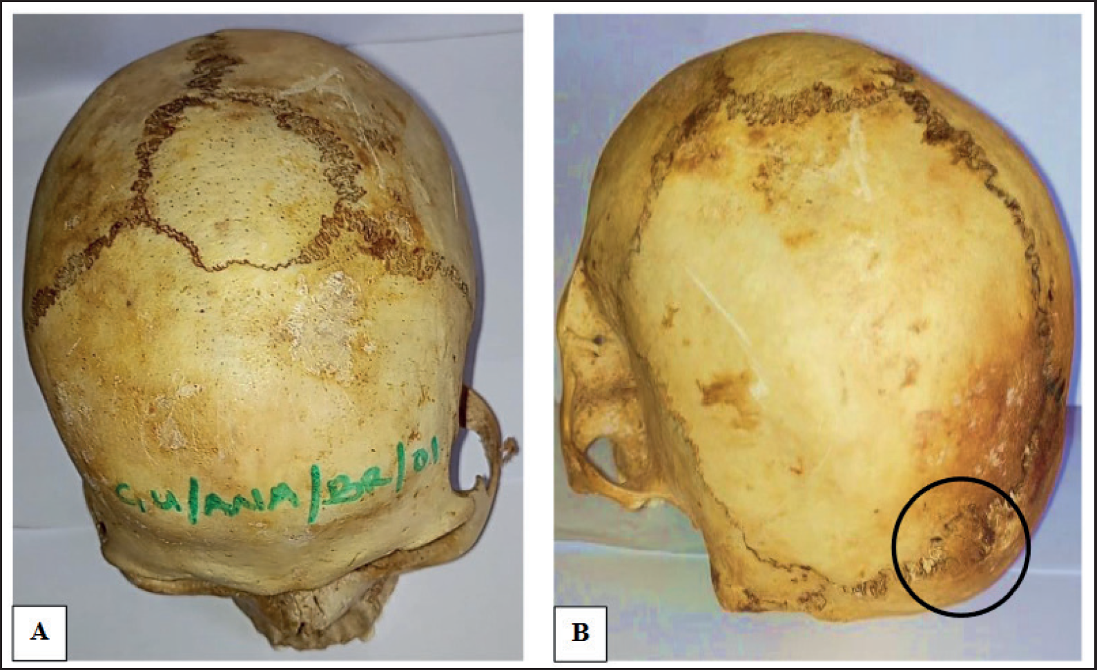
            Figure 1.

            Showing various sutural bones in the skull; (A) bregmatic sutural bone, (B) sutural bone along lambdoid suture.

            Bregma is the location in the skull where sagittal and coronal sutures meet; it is represented by the anterior median fontanelle in the fetal life which obliterates by the 18th postnatal month. The presence of the bregmatic sutural bone was suggested to be a result of an abnormal ossification center in the fibrous membrane at the anterior median fontanelle [2,14]. Bregma is an essential clinical and surgical landmark, so it was proposed that the bregmatic sutural bone be called median frontoparietal sutural bone [2]. Bregmatic sutural bone could be misdiagnosed for skull fractures; hence this reported anatomical variation present at the bregma of the skull forms knowledge that may be of importance to radiologists, orthopedic surgeons, neurosurgeons, and other related professions in their diagnoses and practices.

            Conclusion

            This report displays the first reported case of human bregmatic sutural bone (sutural bone at bregma of the skull) in Nigeria, Africa, and one of the few reported cases across continents of the globe. This supports the possibility of variations in ossification centers and skull matrix; hence, provides knowledge that can aid neurosurgeons, orthopedics, radiologist, anatomists, and other medical/health professionals in their diagnoses and practices.

            What is new?

            The human bregmatic sutural bone is an unusual variation of the human skull that can be misdiagnosed as a skull fracture. The knowledge of this case report reveals and supports the possibility of this anatomical variation, which is of vitality to neurosurgeons, orthopedics, anatomist, anthropologists, and forensic medical professionals in their diagnoses and practices. Although only a few cases have been reported across the continent about the human bregmatic sutural bone. This is the first reported case in Africa.

            Conflict of interests

            The authors declare that there is no conflict of interest regarding the publication of this article.

            Funding

            None.

            Consent for publication

            Not applicable.

            Ethical approval

            Ethical approval is not required at our institution to publish an anonymous case report.

            References

            1. Barberini F, Bruner E, Cartolari R, Franchitto G, Heyn R, Ricci F, et al.. An unusually-wide human bregmatic wormian bone: anatomy, tomographic description, and possible significance. Surg Radiol Anat. 2008. Vol. 30(8):683–7. [Cross Ref]

            2. Hussain Saheb S, Mavishetter GF, Thomas ST, Muralidhar P. Unusual sutural bone at bregma-A case report. Biomed Res. 2010. Vol. 21(4):369–70

            3. Rakesh G, Nema U, Zaidi SHH, Alok SK. Incidence of bregmatic bone in north Indian skulls: incidence of bregmatic bone in north Indian skulls. Natl J Integr Res Med [Internet]. 2013. 2023 Jan. 18Vol. 4(2):79–81

            4. Edwards B, Wang JM, Iwanaga J, Luviano J, Loukas M, Oskouian RJ, et al.. Hiding within the cracks: case report of rare sutural bone found at the nasion. Cureus. 2017. Vol. 9(6):e1333[Cross Ref]

            5. Romero-Reverón R, Arráez-Aybar LA. Sutural bones: a literature review. Anatomy. 2019. Vol. 13(1):61–5. [Cross Ref]

            6. Ratnaningrum SD. Identification of sutural bones in Indonesian skulls. Transl Res Anat. 2020. Vol. 18:100061[Cross Ref]

            7. Noback CR, Ruggiero DA, Strominger NL, Demarest RJ. The human nervous system: structure and function. 2005. New York, NY: Springer Science & Business Media.

            8. Reveron RR. Anatomical classification of sutural bones. MOJ Anat Physiol. 2017. Vol. 3(4):130–1. [Cross Ref]

            9. `Guerrero Jazo FJ, Tovar Spinoza Z, Esparza Gutiérrez SV, Hernández Chávez E, Monroy Rizo EJ. Rupture or traumatic dislocation of cranial suture on infants, involvement of the lambdoidal accessory bones (Wormian bones): case presentation and analysis. Childs Nerv Syst. 2019. Vol. 35(11):2233–36. [Cross Ref]

            10. Johal J, Iwanaga J, Loukas M, Tubbs RS. Anterior fontanelle wormian bone/fontanellar bone: a review of this rare anomaly with case illustration. Cureus. 2017. Vol. 9(7):e1443[Cross Ref]

            11. Rudie L, Lewis PJ. The first record of wormian bones in lizards. Afr J Herpetol. 2018. Vol. 67(2):127–31. [Cross Ref]

            12. Manjunath KY. Supernumerary bones in the walls of the bony orbit. People J Sci Res. 2013. Vol. 6:7–12

            13. Mahajan R, Tuli A, Raheja S, Tomer S, Beri S. Morphometric study of sutural bones of orbit with their clinical implications. Int J Clin Exp Res Med. 2018. Vol. 2:29–36. [Cross Ref]

            14. Ferruh Y, Hulusi E, Zehra A. A Study on the interparietal bone in man. Turk J Med Sci. 1998. Vol. 28:505–9

            Summary of the case

            1 Patient (gender, age) Cadaver specimen (male)
            2 Final diagnosis Not applicable
            3 Symptoms Not applicable
            4 Medications Not applicable
            5 Procedure Maceration
            6 Specialty Anatomy, anatomical variation

            Author and article information

            Journal
            European Journal of Medical Case Reports
            EJMCR
            Discover STM Publishing Ltd.
            2520-4998
            28 February 2023
            : 7
            : 2
            : 27-29
            Affiliations
            [1 ]Department of Anatomy, College of Medicine and Health Sciences, Gregory University Uturu, Uturu, Nigeria
            [2 ]Department of Anatomy, College of Medicine, University of Port Harcourt, Port Harcourt, Nigeria
            [3 ]Department of Physiology, College of Medicine and Health Sciences, Gregory University Uturu, Uturu, Nigeria
            Author notes
            [* ] Correspondence to: Osah Martins Onwuka Department of Physiology, College of Medicine and Health Sciences, Gregory University Uturu, Uturu, Nigeria. osahmartinz@ 123456gmail.com
            Article
            ejmcr-7-27
            10.24911/ejmcr/173-1654940319
            5c4824d6-4123-4bf1-8601-be9e6538141c
            © Nkechi Clara Nwosu, Loveday Ese Oghenemavwe, Osah Martins Onwuka

            This is an open access article distributed in accordance with the Creative Commons Attribution (CC BY 4.0) license: https://creativecommons.org/licenses/by/4.0/) which permits any use, Share — copy and redistribute the material in any medium or format, Adapt — remix, transform, and build upon the material for any purpose, as long as the authors and the original source are properly cited.

            History
            : 11 June 2022
            : 06 January 2023
            Categories
            CASE REPORT

            sutural bone,Bregma,skull,wormian bone,anatomical variation

            Comments

            Comment on this article