Background
The management of ureteric calculi depends on the size and site of the calculus with the likelihood spontaneous passage of calculus depending on the size of the calculus. Usually, small ureteric stones are likely to pass out; however, larger stones that are more than 1 cm in size are less likely to pass out spontaneously [1]. In the modern era, endourological procedures are the preferred treatment modalities for the management of urinary lithiasis with a prevailing dilemma continuing to exist in the management of larger stones. Here we report a case of large ureteric calculus of 3.5 × 2 cm and weighing 35 g, which was successfully managed by laparoscopic ureterolithotomy (LUL).
Case Presentation
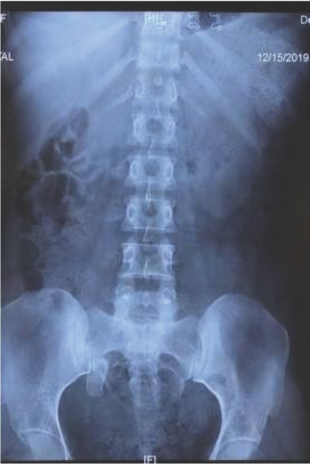
A 30-years-old female with no known comorbidities/prior surgery presented to our outpatient clinic with complaints of intermittent right flank pain and dysuria which was unrelieved for the past 4 months. Physical examination, metabolic work-up, and urine analysis reports were unremarkable. X-ray kidney ureter bladder (KUB) confirmed the presence of a large distal ureteric calculus on the right side (Figure 1), with the Intravenous urogram revealing dilated upper ureter and gross hydronephrosis of the right kidney with poor function (Figure 2). The diethylenetriamine pentaacetic acid radionucleotide renal scans confirmed that the relative function of the right kidney was 11.19% with a glomerular filtration rate (GFR) of 7.59 ml/ minutes, while the opposite kidney had a relative function and GFR of 88.81% and 60.26 ml/minutes, respectively. The patient underwent right LUL with intracorporeal suturing with 3-0 Vicryl(TM) using a modified three-port technique and Double J stenting using a 10-mm umbilical port and two 5-mm ports made at the right sub-costal region and at the level of anterior superior iliac spine in the mid-clavicular line (Figure 3) and a large stone of 3.5 × 2 cm was retrieved (Figure 4). Ureteral reimplantation was not considered necessary in the present case as there was no evidence of any trauma/injury to the ureterovesical junction confirmed by the uneventful smooth placement of the Double J stent after stone extraction, virtually ruling out any possibility that distal ureteral strictures that was confirmed on follow-up. The patient’s postoperative period was uneventful and she is currently doing well, with the follow-up renal dynamic scan imaging at 1 year revealing minor improvements in the function of the right kidney with relative function and GFR of 14.42% and 10.6 ml/minutes, respectively. Fourier transform spectroscopic quantitative stone analysis suggested 80% monohydrate and 20% dihydrate calcium oxalate stones.

Preoperative plain KUB film showing right large opacity in line of the right distal ureter.
Discussion
Giant ureteric calculus is defined as a stone weighing 50 g or measuring 5 centimeters in its greatest dimension. The heaviest and the longest ureter stone reported in literature till date were of 286 g by Mayer et al. [2] and 21.5 cm in length by Taylor et al. [3]. A giant or large ureteric calculus may often occur due to the associated anatomical urinary tract malformations (megaureter, ureterocele, etc.), either alone or with metabolic abnormalities which may predispose the development of massive giant ureteric stones; however, they may occur without the same being detected [4,5]. Some calculi may remain silent or be minimally symptomatic, resulting in partial or even complete loss of renal function at time of presentation, as in this case [6]. While current guidelines exist for the management of smaller ureteric calculi, there exist dilemmas with respect to an ideal surgical modality for significantly larger/giant ureteric calculi since they are unlikely to be cleared by Shock wave lithotripsy (SWL) or endourological procedures due to their higher stone burden and hardness that may necessitate multiple surgical procedures causing a significant financial burden/higher morbidity and a longer hospital stay. The modified technique of LUL used included the use of three ports in place of four ports, modified intracorporeal suturing using barbed sutures, and avoidance of ureteric reimplant. LUL, being a minimally invasive approach, naturally emerges as the ideal therapeutic surgical modality of choice for the management of such a large ureteric calculus as it has the combined advantages of being a single-stage definitive minimally invasive procedure with minimal postoperative pain/lower morbidity, rapid convalescence/shorter hospital stay, and excellent stone clearance rates without any significant long-term sequel [7]. Despite the advances in SWL and/or endourological procedures, the management of giant ureteric stones is often surgical [4].
| NO | AUTHOR | SALIENT CASE DETAILS | MANAGEMENT |
|---|---|---|---|
| 1. | Natami et al. [8], IMCRJ | 106 g, Left DGUC, 32 years/M | Combined-(Endoscopic RIRS +Open Ul) |
| 2. | Maranna et al. [9], Int Surg J | DGUC 4.5 cm | Open Ul |
| 3 | Vaddi et al. [10], Urol | 16 cm, Assoc Bladder exstrophy | Open Ul |
| 4 | Lal et al. [11], Int J Res Med Sci | DGUC, 10.5 cm, 49 g. | Open Ul |
| 5 | Sarikaya et al. [12], Case Rep Med | DGUC-11.5 cm size. | Open Ul |
| 6 | Rathod et al. [13], IJU | 11 cm, 40 g, 35/F, Rt NFK | Lap Ul |
| 7 | Barry et al. [14], Br J Med Surg Urol | Bilateral DGUC,12.8 g, Duplex system | Open Ul |
| 8 | Jeong et al. [15], Clin Nephrol | 6 × 2cm, Rt atrophic renal unit | T/P Lap Ul |
| 9 | Mak et al. [16], J Surg Case Rpt. | Silent DGUC, 7 and 3 cm, 7/M. | Open Ul |
| 10 | Jouini et al. [4], Prog Urol | 2 Cases DGUC | Open Ul |
| 11 | Kim et al. [17], Korean J Urol | Hen-egg sized DGUC, Lt NFY, 54/F | Open uretronephrectomy |
| 11 | Present case | DGUC, 3.5 cm, 38 g | Lap Ul |
DGUC = Distal Giant Ureteric Calculi; UL = Ureterolithotomy; RIRS = Retrograde Intra-renal surgery; NFK = Non Functioning Kidney; T/P Lap UL = Transperitoneal laparoscopic ureterolithotomy.
Conclusion
LUL holds the key to the management of select uncomplicated large ureteric calculi with the advantage of being minimally invasive accompanied by good clearance rates thereby acting as a bridge to combine the positives of both endourological and open surgical procedures.
What is new?
Endourology provides an exciting option for the management of urinary lithiasis, but this surgical branch comes with the inherent downsides of requirement of multiple settings in larger stones, incomplete clearances, and increased patient costs owing to the mentioned disadvantages. In this article, we present the novel but less used technique of LUL which combines the advantages of ensuring complete clearance and being minimally invasive in the management of larger ureteric stones. The void in the knowledge of practicing laparoscopy in distal larger giant ureteric stones is briefly depicted in Table 1, where a majority of the large ureteric stones were managed by the outdated open technique rather than laparoscopy. This gap in the use of minimally invasive surgical management of distal giant/large ureteric calculi by surgeons is expected to be bridged by the present article by way of adding to the scarce literature on the subject.


