Background
Chest trauma is one of the major causes of mortality in road traffic accidents. Blunt chest trauma may result in a variety of skeletal and visceral injury due to crush or blast impact while hemothorax following blunt chest trauma commonly resulted from multiple rib fractures and diaphragmatic injury [1]. The incidence of delayed hemothorax from blunt chest trauma ranges from 5.0% to 7.4%. Delayed massive hemothorax complicating blunt chest injury, on the other hand is exceedingly rare in clinical setting and is potentially life threatening [2–4]. A massive hemothorax is defined as pleural blood drainage of equal or more than 1,500 ml or with continuous bleeding at a rate of 200 ml/hour for at least four consecutive hours [4]. Studies have found that delayed hemothorax following blunt chest trauma typically present within the first week post-injury, while others describe up to 44 days in delay of presentation [5]. This prolonged presentation may be attributed to the slow seeping of blood from the smaller blood vessel that might have been injured at the point of impact and the oblivion of the condition causes no limitation to the physical movement of the patient, further extending the physical damage of the initial injury. Generally, cases of massive hemothoraces encountered in clinical settings warrant admission immediately following trauma for continuous observation and management. However, in cases of delayed hemothorax in hemodynamically stable patients, clinical observation over a certain period of time is commonly practiced, rarely requiring emergency surgery [1].
We present a case of an elderly gentleman who sustained multiple rib fractures from alleged motor-vehicle accident. Despite initial in-patient monitoring, he presented to us again with overt massive hemothorax.
Case Presentation
This is a case of a 65-year old gentleman with no known past medical illness with alleged motor-vehicle accident (motorbike vs. motorbike). He sustained right second to eigth rib fractures and a comminuted fracture of the right scapula (Figure 1A). He was otherwise alert on presentation and his hemodynamic status was stable.
Immediately following injury, he was monitored in-patient with strict bed rest for 5 days for clinical red flags. His vital signs remained stable throughout hospitalization and he was discharged well thereafter with advice. No further radiological imaging was done during this period.
Unfortunately, he presented back to us 2 days after his hospital discharge [on the seventh day post-motor-vehicle accident (MVA)] with sudden onset of right-sided chest pain and shortness of breath. He was otherwise alert and conscious. His blood pressure was 112/69 mmHg, he was tachycardic (Pulse rate: 138 bpm), afebrile, mildly tachypneic (Respiratory rate: 20 breaths/minutes) and his SpO2 reading was 90% under room air.
The right chest wall was dull to percussion and there was reduced air entry all over the right lung field. Urgent chest X-ray revealed right lung opacity up to the middle zone with blunting of right costophrenic angle (Figure 1B). Right thoracostomy tube was inserted (Figure 1C), draining 800 ml of frank blood upon insertion and a total of 1,250 ml within the next 12 hours.
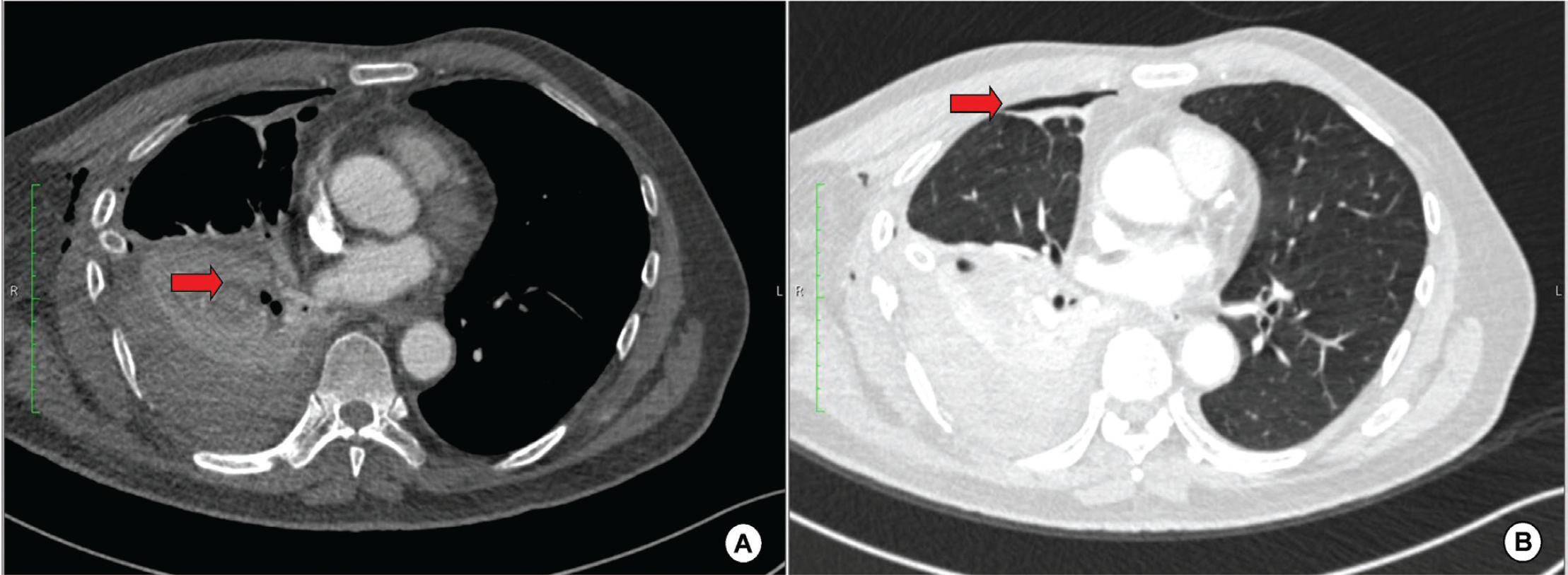
Urgent computed tomography (CT) angiogram of the thorax revealed signs of pulmonary laceration at the right lower lobe (Figure 2A) with multiple rib fractures and right comminuted scapular fracture. Minimal right pneumothorax was also noted (Figure 2B).
The complex rib fractures include multiple two-part fractures of the right second to eighth ribs and bilateral first rib fractures. Packed cell was transfused accordingly and tranexamic acid 1 g 8-hourly was given in the emergency setting. Adequate analgesia was addressed throughout hospitalization. The patient was subsequently transferred to the cardiothoracic center for definitive management.
Discussion
We report a case of delayed massive hemothorax due to pulmonary laceration and multiple rib fractures caused by a blunt thoracic trauma following moderate impact motor-vehicle accident.
With regards to trauma cases in emergency setting, timely localization of vascular, skeletal, or airway injury following trauma allow early diagnosis, and represent the cornerstone of medical-surgical treatment and planning of clinical management [6]. CT scan plays a key role in the diagnostics of chest trauma with a considerable impact on the ensuing therapeutic decisions.
Delayed massive hemothorax may result from intercostal or phrenic artery tearing, laceration of the diaphragm, or fractured ribs [2]. However, our patient presented to us with signs of acute respiratory failure due to massive hemothorax resulting from pulmonary laceration and multiple complex rib fractures.
The possibility of delayed sequelae following blunt chest trauma should continually be communicated to the patient and the family to encourage vigilance and monitoring even after being discharge home from the initial hospital monitoring, as the delay between the trauma and the onset of presentation of sequelae typically vary from 18 hours to 11 days [7]. In our case, despite being initially admitted under close monitoring for the first 5 days following injury with no red flags, he came in 2 days later with abrupt clinical deterioration.

Chest radiographs. (A) The first chest radiograph following MVA showing right second to eigth rib fractures. (B) Chest radiograph taken at Day-7 post-trauma showing homogenous opacity up to the right middle zone. (C) Chest radiograph post-chest tube insertion at the Emergency Department showing signs of clearing up of the right lung field.

CT thorax. (A) Multiple areas of hypodense lesions (red arrow) in the right lower lobe suggestive of pulmonary laceration. (B) Minimal right pneumothorax (red arrow).
It may be helpful to stratify the patients at higher risk to develop delayed complication with hemothorax, particularly in patients with sharp edges broken ribs [8,9]. In such cases, earlier CT scans may alert the clinicians of possible complications that could be anticipated. In our case, no detailed radiological assessment was done during the initial in-patient monitoring.
Additionally, in cases of minimally displaced rib fractures, the sharp edges of the broken ribs may still injure the surrounding viscera, particularly with continuous physical movement, hence patient education and counseling upon discharge is of utmost importance.
Thoracocentesis and chest tube placement are often sufficient to manage the clinical symptoms and save the lives of many thoracic trauma patients affected by lung or airway injuries [6]. Despite rarely requiring emergency surgery, delayed massive hemothorax is still potentially life threatening [10].
Conclusion
All patients with blunt chest trauma should be informed of the need for a close observation upon admission, even if the fractured ribs are not severely displaced. Fractures involving three or more sequential ribs and a flail chest are considered as complex thoracic injuries and are frequently associated with a significant degree of hemothorax.
Rapid and accurate diagnoses with timely intervention are keys to success in the management of trauma patients with complications. Herein, we report this case to share our valuable experience and to encourage our colleagues in emergency settings and physicians worldwide to practice vigilance when encountering patients with blunt thoracic trauma.

