Background
Extra-nodal diffuse large B-cell lymphoma (DLBCL) is a heterogeneous group of uncommon neoplasms, arising in almost every body site such as intestine, breast, bone, liver, skin, lung, testis, and central nervous system [1]. Muscle involvement of DLBCL is, especially, rare [2,3]. Typical clinical manifestation of cutaneous DLBCL includes patches, plaques and non-ulcerated solitary nodules, single or multiple, with firm consistency, and low rate of extra-cutaneous dissemination [4,5]. The management of cutaneous DLBCL is carried out with multi-modality treatment strategy, which may include surgical resection, chemotherapy, radiotherapy, antibiotics, immunotherapy, corticosteroids, etc. [6] The prognosis of cutaneous DLBCL is generally more favorable than their nodal counterparts, where the type of tumor and degree of cutaneous involvement are considered as the important prognostic factors [7–9].
Herein, we present a case of diffuse large B-cell lymphoma of the left leg involving the gastrocnemius muscles and assess the response of our treatment protocol (i.e., neoadjuvant chemotherapy, total tumor resection followed by conventional radiotherapy). The patient is being followed up regularly and has remained disease-free for over 1.5 years.
Case Presentation
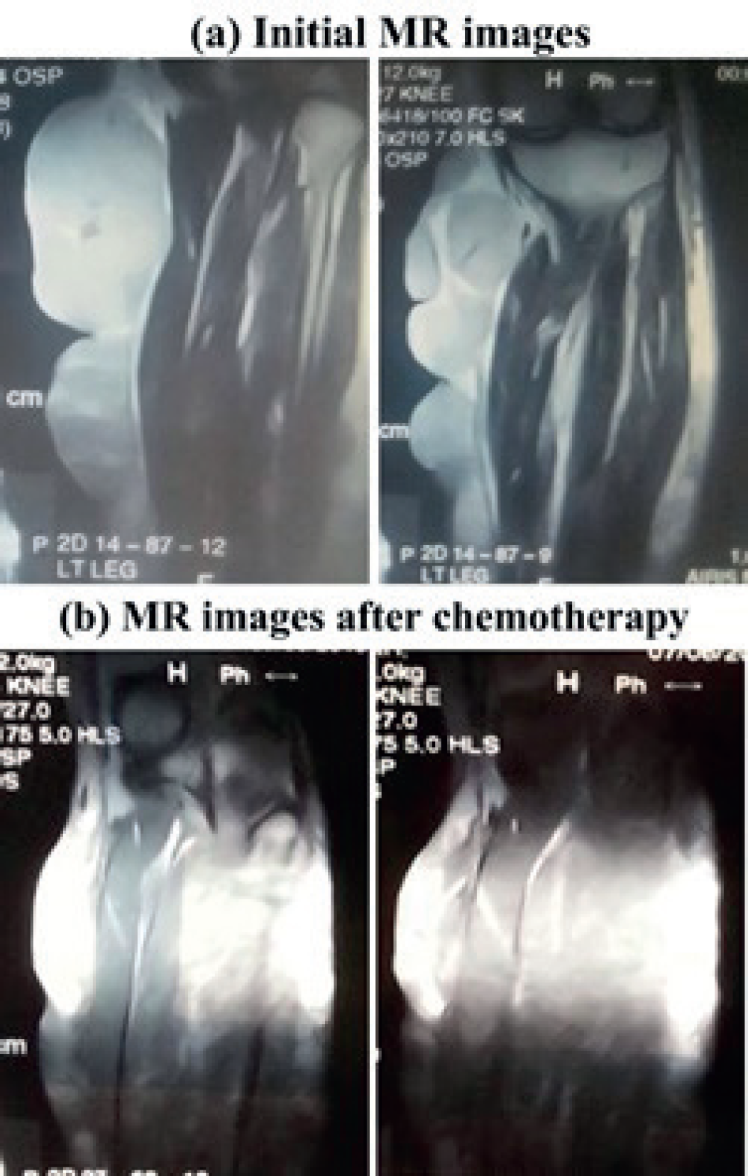
A 60-year-old male presented with large swelling of left leg for one year. Clinically, the patient had multi-lobulated mass on the left leg, below the knee joint (Figure 1a). Magnetic resonance (MR) images of the left leg showed abnormal, heterogeneous signal mass (size = 18 × 14 cm) involving the subcutaneous tissues and the overlaying skin (Figure 2a). The mass also involved the gastrocnemius muscles. Surgical resection of the mass was not possible. The patient underwent incisional biopsy of the mass. Histological examination revealed a lymphoid neoplasm composed of large tumor cells arranged in a discohesive fashion with scattered lymphocytes in the background. The individual neoplastic cells had rounded to vesicular nuclei, conspicuous nucleoli, and appreciable amount of cytoplasm. Numerous mitosis was also seen. Immunohistochemical staining (IHC) showed CD20 positivity in tumor cells. The proliferative index Ki 67 (Mib-1) was more than 80%. These features favored cutaneous DLBCL of the left calf.
The patient was planned for neoadjuvant chemotherapy. Specifically, the patient received six courses of CHOP regimen (i.e., cyclophosphamide; 750 mg/m2, doxorubicin; 50 mg/m2, vincristine; 1.4 mg/m2, and prednisone;100 mg/ m2). The tumor response to the chemotherapy was very good, with a significant reduction in the tumor size, both clinically (Figure 1b) and radiologically as demonstrated by the reassessment MR images (Figure 2b). Afterward, the patient underwent complete tumor resection, with a loss of skin after surgery. After 3 months, skin grafting was performed (Figure 1c). At this stage, histopathology was unremarkable. Specifically, the excised sections revealed unremarkable epidermis and underlying dermis. The subcutaneous tissue showed areas of fibrosis, edema, and scattered blood vessels. The underlying skeletal muscle tissue was also unremarkable. IHC staining showed reactive pattern for CD20, CD3, and PAX-5. Overall, no residual lymphoma was identified on extensive sampling. After 4 months of the skin grafting, the patient received radiotherapy (36Gy, standard fractionation) to the tumor bed. Presently, the patient is being followed up regularly and has remained disease-free for over 1.5 years. Written informed consent for publication of this case report has been obtained from the patient.

Image of the patient presented with DLBCL of the leg; (a) photo at presentation illustrating large, multi-lobulated mass on the left leg, below the knee joint right, (b) photo after six course of neoadjuvant chemotherapy showing significant reduction in the tumor size, and (c) photo after surgical resection demonstrating no residual tumor.

MR images of the left leg: (a) initial images at the presentation illustrating abnormal, heterogeneous signal mass (size = 18 × 14 cm) involving the sub cutaneous tissues and the overlaying skin, and (b) images after six course of neoadjuvant chemotherapy showing significant reduction in the tumor size.
Discussion
DLBCL is an aggressive form of non-Hodgkins lymphoma, predominantly occurring in older age (i.e., > 60 years), with or without “B” symptoms, including fever, night sweats and weight loss [10]. The most common site for extra-nodal presentation is the gastrointestinal tract or bone marrow [6]; nevertheless, it also manifests at less common sites, such as the skeletal muscle or testis [2,11]. When the skeletal muscle is involved, it is predominantly the upper arm muscles and glutei. The lower skeletal muscle involvement, as reported here, is very uncommon.
The primary therapeutic option for DLBCL tumors is the complete surgical resection that markedly improves the prognosis [4,6]. However, complete surgical resection occasionally appears challenging due to the involvement of vital structures by the aggressive tumor. Alternatively, the patient may be treated with neoadjuvant chemotherapy to reduce the tumor burden to the point where complete resection becomes possible, as seen in the present case.
The recommended chemotherapy for DLBCL is the CHOP regimen (i.e., cyclophosphamide, doxorubicin, vincristine, and prednisone). Sometimes, this chemotherapy is coupled with immunotherapy (i.e., rituximab) [12]. We also followed the same chemotherapy protocol (i.e., CHOP) for our DLBCL patient, as the patient was non-affording for rituximab and observed markedly good response in terms of tumor size reduction. To further diminish the risk of recurrence, the patient received radiotherapy to the tumor bed. With this management protocol, it has been 1.5 years down the road without any recurrence or metastatic disease.
Conclusion
DLBCL of the leg is a rare malignancy which is typically seen in older age. Herein, we presented a case of DLBCL of the left leg. The patient showed marked improvement in terms of tumor size reduction with CHOP regimen-based neoadjuvant chemotherapy. Afterward, complete surgical resection of the tumor was carried out, followed by conventional radiotherapy (i.e., 36 Gy/18 fractions). The patient is on regular follow up for more than 1.5 years, without any disease recurrence or metastasis. This case report demonstrates that DLBCL is an aggressive malignancy and responds promisingly to multi-modality treatment strategy (i.e., chemotherapy, surgery, and radiotherapy).


