Background
Adrenal myelolipoma is characterized by a certain type of lesion, benign tumor (2%–4% of adrenal type) [1] and considered the major type of adrenal incidentalomas [2]. These types of tumor sometimes known as asymptomatic, unilateral, or bilateral that might cause symptoms [3]. A stimulus (necrosis, stress or infection, and inflammation) was found to increase the prevalence of adrenocortical metaplasia that considered the most type of theories [4]. Adrenal myelolipoma has a very tiny diameter size (< 5 mm). Performing surgery for cases was rare, while the rate of detection increased recently due to the use of imaging modalities [5]. Adrenal myelolipoma also named incidentaloma when the cases were detected as asymptomatic incidental imaging findings [6]. It commonly seen in a population of age that ranged between 40 and 60 years and similarly distributed between males and females [5]. This study reported two patients with incidentally detected myelolipoma and was selected for surgery according to their clinical symptoms. Both patients recovered quickly without postoperative complications.
Case Presentation
A 58-year-old male was presented with discomfort in the right upper abdomen and a palpable mass in physical examination caused by a giant adrenal myelolipoma. Abdominal ultrasound showed a large hyperechoic mass in the right upper quadrant measuring 14 cm. Routine hematological parameters were normal. Serum catecholamine, cortisol, and urinary, vanillylmandelic acid were within a normal range. Contrast-enhanced computed tomography (CT) scan of the abdomen revealed a well-defined, large, round lesion in the right adrenal region measuring about 14 cm with heterogeneous attenuation suggested the possibility of myelolipoma. The diagnosis of symptomatic myelolipoma was made and the patient was scheduled for trans-abdominal right adrenalectomy through the right subcostal incision and his postoperative course was uneventful. The histopathological evaluation of the mass revealed a characteristic mixture of mature adipose tissue with a hematopoietic element without malignancy, confirmed the initial diagnosis of adrenal myelolipoma.
Another case of a 43-year-old female with a known sickle cell anemia presented with weight loss and chronic abdominal pain in the right upper abdomen without a history of fever. Physical examination revealed a cachectic patient (body mass index was 18) with known splenomegaly and a palpable mass in the right upper abdomen. The abdominal ultrasound examination showed splenomegaly as well as a hyperechoic mass in the right upper quadrant measuring 8 cm in diameter. Routine laboratory parameters were normal except for an elevated iron-level secondary to previous multiple transfusion. Serum catecholamine, cortisol, plasma-metanephrine, and urinary vanillylmandelic acid levels were within a normal range. The patient was scheduled for endoscopic right adrenalectomy through a minimally invasive retroperitoneal approach. The postoperative course was uneventful. The histopathological evaluation of the mass revealed a characteristic mixture of mature adipose tissue with a hematopoietic element without malignancy, confirmed the initial diagnosis of adrenal myelolipoma.
CT appearance of myelolipoma showed the heterogeneous mass covering upper right retroperitoneal space with variable central and peripheral attenuation. Homogenous, sharply circumscribed, the hyperechoic tumor was adjacent to the right kidney (Figure 1).
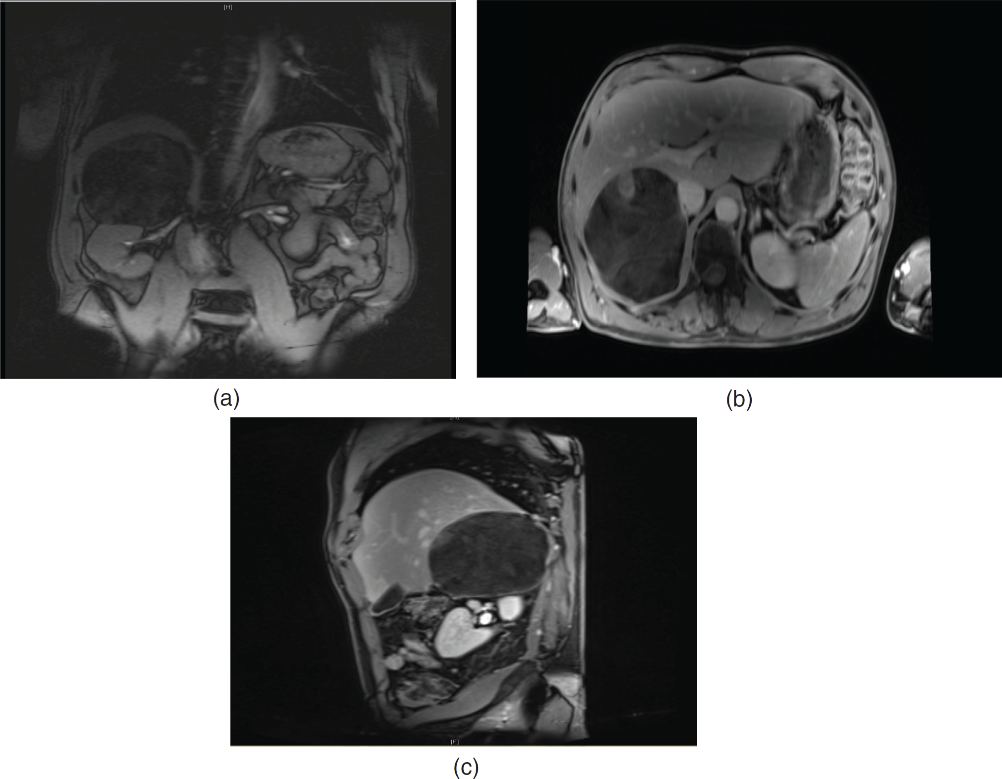
Magnetic resonance imaging (MRI) appearance of myelolipoma showed a large hyperintense right adrenal mass with central hypointense areas as well as signal drop suggested fat content in the mass (Figure 2A–C).
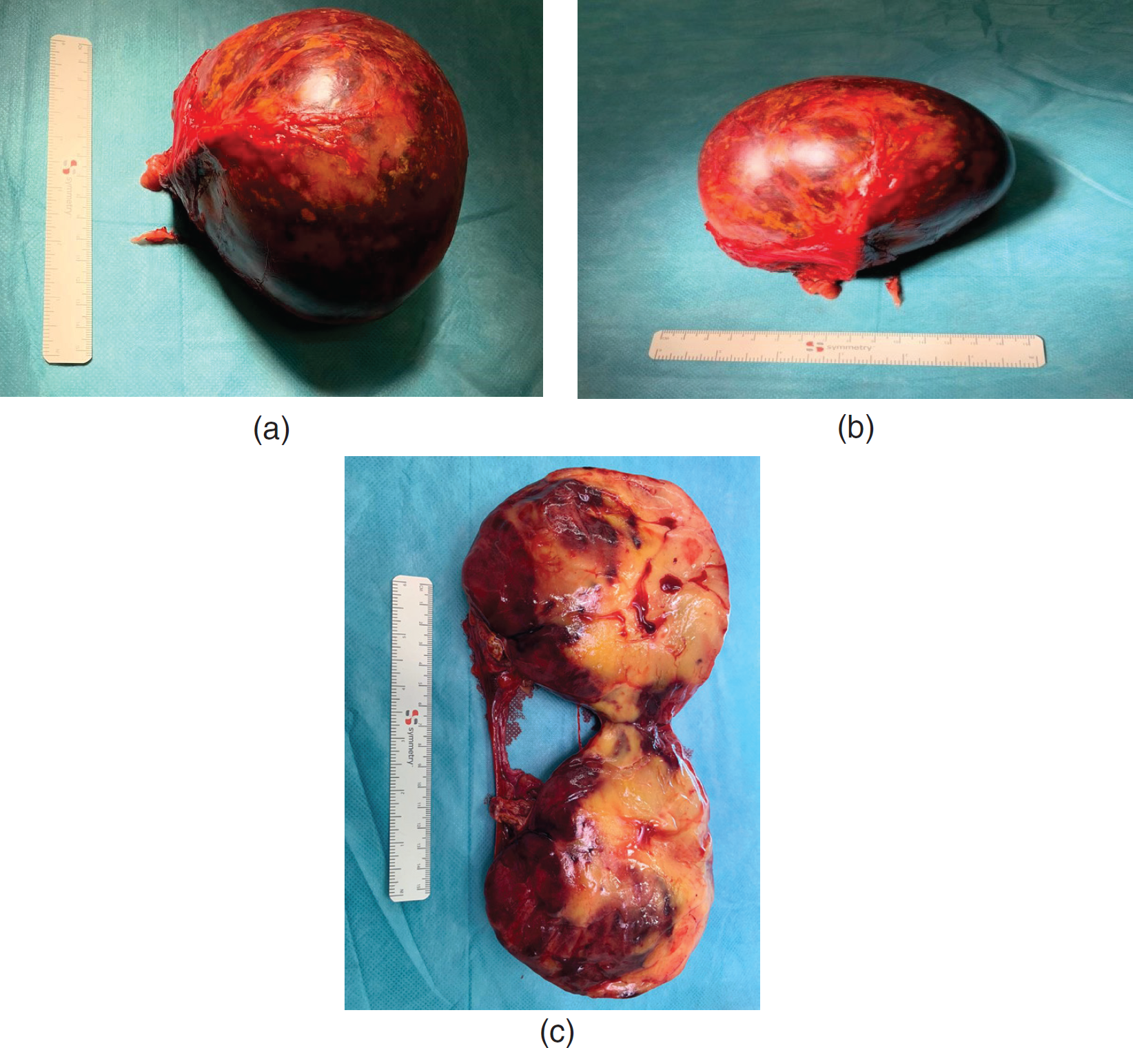
Gross appearance of an Adrenal myelolipoma showed a large capsulated mass with adipose tissue and hemorrhagic areas within it (Figure 3A–C).
Both symptomatic and hormone active myelolipomas were indicated for surgery (Figure 4).
Furthermore, management approaches for adrenal incidentalomas were also reviewed (Figure 5).
Discussion
Adrenal myelolipomas are a type of benign tumors of hematopoietic that are non-functional and considered as a mature adipose tissue. It is usually small, mono-lateral, and asymptomatic tumor; it represents almost 1.9% of adrenal incidentalomas [7]. They are also called “incidentalomas” since their diagnosis is based on autopsy or imaging modalities which are performed for reasons usually unrelated to adrenal diseases. The incidence ranges from 0.08% to 0.4%, and less than 300 cases were reported in the literature before 2000 [8]. However, their prevalence appears to be increased by up to 10%, due to the increased use of noninvasive and enhanced imaging techniques [9].
In this case report, both patients suffered from symptoms caused by the mass effect of their myelolipomas. The patients were scheduled for conventional trans-abdominal and minimal-invasive retroperitoneal surgery according to their tumor size. Conventional approaches, including midline, subcostal, lumbar, or posterior access laparotomy, had been described in the literature. An extraperitoneal approach was preferable as it led to quicker recovery of the patient and lesser postoperative complications [10]. After excision, adrenal myelolipoma generally does not recur, with recurrence-free survival rates of up to 12 years being reported. There was no evidence of malignant transformation in the literature. German guidelines recommended 6 cm diameter as a limit to minimally invasive surgery [11]. In the institutions with expertise in adrenal surgery, benign tumors below 10 cm diameter could be resected minimally invasive by retroperitoneal access, first described by Liu et al. [12]. In minimally invasive adrenal surgery, malignancy has to be excluded preoperatively, because retrieved specimen had to be morcellated intracorporeally. A variety of differential diagnosis of adrenal masses has to be noted, important was to rule out malignancy before the surgical intervention.
In the present study, after excluding adrenal metastases and adrenocortical carcinoma by its low proportion of fatty tissue on CT, there was still retroperitoneal liposarcoma and renal angiomyolipoma to be considered. Retroperitoneal liposarcoma is a slow-growing ill-defined tumor of the retroperitoneal region which is composed of adipocytes and radiologically appears similar to an adrenal myelolipoma with an invasion of the adjacent structure. Renal angiomyolipomas are tumors arising from the kidney, they consist of fat, blood vessels, and muscle cells. These are large, heterogeneous appearing tumors that could invade through the renal capsule. Image-guided biopsy for diagnostic confirmation was useful and safe. However, care must be taken to avoid the risk of malignant, seeding, rupture, and bleeding. Twenty two cases had been reported in the literature [13] with bleeding complication, especially intratumoral hemorrhage. Characteristics included sudden onset of pain, male dominance, right-sided predilection, and mean tumor size of 11.7 cm (range 4–20.5).

(A) MRI appearance of myelolipoma. (B) MRI appearance of myelolipoma. (C) MRI appearance of myelolipoma.

(A) Gross appearance of an adrenal myelolipomas. (B) Gross appearance of an adrenal myelolipomas. (C) Gross appearance of an adrenal myelolipomas.
Therefore, it could be said that the management approach should be individualized based on the presence of symptoms and the size of the lesion. Asymptomatic tumors could be usually followed. The surgery is indicated by symptomatic tumors, the presence of signs of a hormone-secreting tumor, or giant myelolipomas due to the risk of intra-tumoral bleeding and rupture. Thus, it was concluded that Adrenal myelolipomas consist of mature fatty tissue combined with hematopoietic tissue, such as myeloid and erythroid cells. Referring to incidentalomas, they are often serendipitously discovered by radiological examination. The presence of macroscopic fat on imaging is characteristic of an adrenal myelolipoma. Surgery is indicated for symptomatic myelolipomas. Asymptomatic tumors could be usually followed, the presence of signs of a hormone-secreting tumor or giant myelolipomas due to risk of intra-tumoral bleeding and rupture. Resection could be performed minimal-invasive up to 10 cm in specialized centers and via open surgery in every other case.
What is new?
Adrenal myelolipomas are a type of benign tumor that is often serendipitously discovered by radiological examination. 2. Asymptomatic tumors could be usually followed, the presence of signs of a hormone-secreting tumor or giant myelolipomas due to risk of intra-tumoral bleeding and rupture 3. Surgery was indicated for symptomatic myelolipomas. Resection could be performed minimal-invasive up to 10 cm in specialized centers and via open surgery in every other case.


