60
views
0
recommends
+1 Recommend
1 collections
    0
    shares
      scite_
      0
      0
      0
      0
      Smart Citations
      0
      0
      0
      0
      Citing PublicationsSupportingMentioningContrasting
      View Citations

      See how this article has been cited at scite.ai

      scite shows how a scientific paper has been cited by providing the context of the citation, a classification describing whether it supports, mentions, or contrasts the cited claim, and a label indicating in which section the citation was made.

       
      • Record: found
      • Abstract: found
      • Article: found
      Is Open Access

      Sarcomatoid carcinoma of maxillary sinus

      Published
      case-report
      Bookmark

            Abstract

            Background:

            Sarcomatoid carcinoma (SC) of paranasal sinuses is a rare variant of squamous cell carcinoma. We present the case of a patient with maxillary sinus tumor projecting into the oral cavity.

            Case Presentation:

            We describe a 79-year-old male who visited the oncology department with complaints of soft tissue growth in the oral cavity. An imaging showed an infiltrating left maxillary antrum mass with locoregional destruction and multiple bone metastases. Histopathology proved this mass as SC. The patient was unfit for surgical resection, so he managed palliatively with radiotherapy.

            Conclusion:

            Being a rare entity, management is debatable, but radical surgery followed by adjuvant radiotherapy confers a better disease control. However, if tumor is unresectable, then radiotherapy alone can be helpful in the palliative management of this tumor.

            Main article text

            Background

            Sarcomatoid carcinoma (SC), also known as spindle cell carcinoma and polypoid squamous cell carcinoma, is a rare variant of squamous cell carcinoma characterized by dysplastic surface squamous epithelium along with an invasive spindle cell element [1]. It can occur at any age from 20 to 90 years with a definite male predilection. The clinical presentation is mostly exophytic and polypoid, but the nodular or endophytic appearance has also been described [1]. SC shows a biphasic histologic appearance with epithelial changes varying from dysplasia to invasive carcinoma and stromal component composed of fusiform or spindle-shaped cells. SC is an extremely rare tumor of the sinonasal tract with only 13 cases reported since 1957 in the literature [2,7].

            Case Presentation

            A 79-year-old male reported to the oncology department with the complaint of soft tissue growth in the left side of the oral cavity for 1 year which was painless but increasing rapidly in size (Figure 1). It was associated with dysphagia. The biopsy showed it as SC. The patient had Eastern Cooperative Oncology Group (ECOG) performance status three on presentation with the bulging left cheek. The patient was nonsmoker, non-diabetic, or hypertensive.

            The patient was investigated with contrast-enhanced Computer Tomography (CT) face and neck that showed a well-lobulated heterogeneously enhancing soft tissue density mass infiltrating left maxillary antrum with the destruction of inferior, anterior, medial, and lateral maxillary wall with the involvement of left-sided hard palate as well as maxillary alveolus (Figure 2). the incidental finding of a mass lesion at the point of confluence of the right and left hepatic ducts with dilated right and left hepatic ducts was made on quadruple-phase CT scan which is highly suspicious for Klatskin’s tumor. Furthermore, biopsy and histopathological confirmation of this lesion was not possible due to the patient’s condition. A biopsy of mass from the oral cavity was done, which showed malignant spindle cell neoplasm. The location of tumor and focal cytokeratin positivity favored SC. The bone scan showed the foci of abnormal radiotracer uptake in manubrium and multiple ribs bilaterally—compatible with skeletal metastases (Figure 3). The rest of the metastatic workup was unremarkable. Hence, the patient was diagnosed as SC of the maxillary sinus with skeletal metastasis.

            The case was discussed in a multidisciplinary meeting, and the patient was referred to an orofacial surgeon, but the surgeon refused surgical resection due to the age and performance status of the patient.

            The patient was given palliative radiotherapy 50G/20fx. Six months post-radiotherapy, patient’s quality of life is better, and he is doing well.

            Discussion

            SC is a rare variant of squamous cell carcinoma characterized by dysplastic surface squamous epithelium along with an invasive spindle cell element [1]. Different authors have different views regarding the histogenesis of SC and have used various terms to describe it. Virchow (1864) first reported it and labeled it as carcinosarcoma, suggesting that it may be a “collision tumor” between carcinoma and sarcoma [3]. Krompecher (1900) proposed an epithelial origin with “dedifferentiation” to a spindle cell morphology and used the term “SC” to describe it. Lane (1957) proposed the term “pseudocarcinoma,” suggesting that it may be a squamous cell carcinoma with an atypical reactive stroma. This multiplicity in nomenclature indicates the complexity of its histogenesis [1].

            It has a wide age of occurrence ranging from 2nd to 9th decade and a mean age during the 5th decade with a predominant male predilection [7]. Although most tumors in the head and neck region occur in the larynx, in the oral cavity, it has a site predilection for the lower lip, tongue, and alveolar ridge or gingiva. Vishwanathan et al., in their study of 103 cases of SC, reported an incidence of 17.5% in the larynx and 63.1% in the oral cavity [4]. In the larynx, true cords and the supraglottic areas are the predominant sites of occurrence with the subglottic area being an unusual location. Pyriform sinuses are the preferred site in the pharynx as are nasal cavity and maxillary antrum in the sinonasal tract [9].

            Because of its rarity, tumor histogenesis, clinical course, and resulting prognosis, its management is debatable [10]. The treatment of choice is radical surgery [46]. Prognosis has been better in patients treated with surgery and adjuvant radiation therapy [6]. Radiotherapy might be administered for the postsurgical palliation of local recurrence. Carcinosarcoma at the maxillary sinus confers a poor survival rate and a high local recurrence rate.
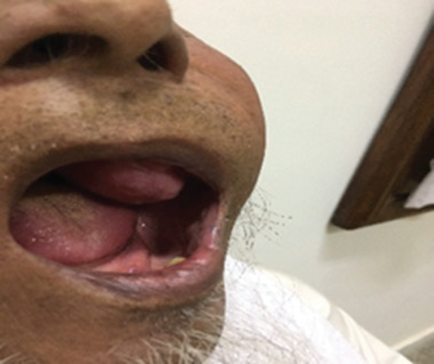
            Figure 1.

            Clinical presentation of the patient. A polypoidal growth hanging into the left side of the oral cavity.
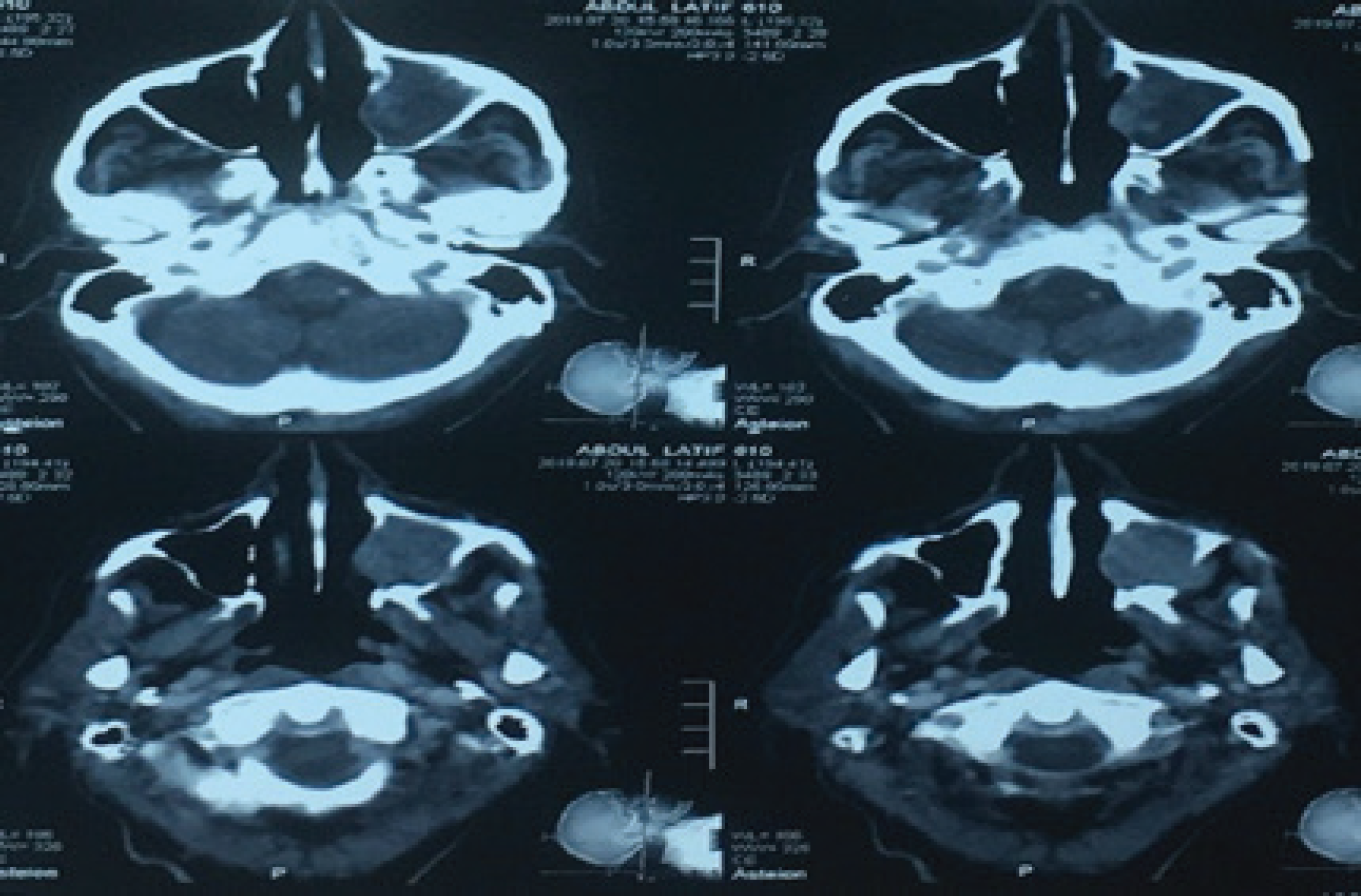
            Figure 2.

            CT manifestation of tumor showing well-lobulated heterogeneously enhancing soft tissue density mass infiltrating left maxillary antrum with the destruction of inferior, anterior, medial, and lateral maxillary wall with the involvement of left-sided hard palate as well as maxillary alveolus.

            Figure 3.

            Bone scan showing metastases bilaterally in ribs and manubrium sterni.

            Conclusion

            Carcinosarcoma of maxillary sinus due to its rarity, tumor histogenesis, clinical course, and resulting prognosis is difficult to manage. Ideally, it should be managed in multidisciplinary meetings via surgical resection and radiotherapy according to the performance status of the patient. The patient can benefit from radiotherapy if this tumor is unresectable.

            What is new?

            SC of the maxillary sinus is a rare tumor with few cases reported, and less is known about its adequate management. Radical surgical resection followed by radiotherapy has been described in the literature as an optimal approach for its management. In the present case, age and poor performance were the factors limiting the optimal management, so radiotherapy alone was delivered to the patient leading to better disease control, quality of life, and manageable adverse effects.

            List of Abbreviations

            ECOG

            Eastern Cooperative Oncology Group

            CT

            Computer Tomography

            SC

            Sarcomatoid Carcinoma

            PNS

            Paranasal sinusses

            Declaration of conflicting interests

            The authors declare that there is no conflict of interests regarding the publication of this case report.

            Funding

            None

            Consent for publication

            Written informed consent was obtained from the patient to publish this case in medical journal.

            Ethical approval

            Ethical approval is not required at our institution for publishing a case report in medical journal.

            References

            1. Mahajan A, Mohanty S, Ghosh S, Urs AB, Khurana N, Gupta S. Sarcomatoid carcinoma of the oral cavity: a diagnostic dilemma. Case Rep Dent. 2017. Vol. 2017:e7495695[Cross Ref]

            2. Hasnaoui J, Anajar S, Tatari M, et al.. Carcinosarcoma of the maxillary sinus: A rare case report. Ann Med Surg (Lond). 2017. Vol. 19:41–4. [Cross Ref]

            3. Basu A, Mondal A. Carcinosarcoma of oral cavity: a unique but rare entity. Sch J Surg. 2018. Vol. 1(1):8–11

            4. Patel TD, Vazquez A, Plitt MA, Baredes S. Eloy JA A case control analysis of survival outcomes in sinonasal carcinosarcoma. Am J Otolaryngol. 2015. Vol. 36(2):200–4. [Cross Ref]

            5. Agarwal SK, Singh S, Sharma S, Lahiri AK. Carcinoma of Hypopharynx a rare entity with unique surgical procedure. Int J Otolaryngol Head Neck Surg. 2013. Vol. 2:259–62. [Cross Ref]

            6. Moon JK, Kim AY. Chang DS Carcinosarcoma of the maxillarysinus. Clin Exp Otorhinolaryngol. 2013. Vol. 6:114–6. [Cross Ref]

            7. Furuta Y, Nojima T, Terakura N, Fukuda S. Inuyama Y A rare case of carcinosarcoma of the maxillary sinus with osteosarcomatous differentiation. Auris Nasus Larynx. 2001. Vol. 28:S127–9. [Cross Ref]

            8. Thompson L, Chang B, Barsky SH. Monoclonal origins ofmalignant mixedtumors (carcinosarcomas):evidence for adivergenthistogenesis. Am J Surg Pathol. 1996. Vol. 20:277–85. [Cross Ref]

            9. Feinmesser R, Wiesel J, Deutsch E, Sela M, Gay I. Carcinosarcoma of the nose and paranasal sinuses-a case report. Rhinology. 1982. Vol. 20(3):167–70

            10. Goellner JR, Devine KD, Weiland LH. Pseudosarcomaofthelarynx. Am J Clin Pathol. 1973. Vol. 59:312–26. [Cross Ref]

            Summary of the case

            1 Patient (gender, age) A 79-year-old male who visited the oncology department with the complaint of soft tissue growth in the oral cavity
            2 Final diagnosis SC left maxillary sinus
            3 Symptoms The patient had a history of mass hanging in the left side of the oral cavity that was painless and associated with dysphagia
            4 Medications None
            5 Clinical procedure Radiotherapy
            6 Specialty Oncology

            Author and article information

            Journal
            European Journal of Medical Case Reports
            EJMCR
            Discover STM Publishing Ltd.
            2520-4998
            30 June 2020
            : 4
            : 6
            : 202-205
            Affiliations
            [1 ]PINUM Cancer Hospital, Faisalabad, Pakistan
            Author notes
            [* ] Correspondence to: Rafshan Sadiq PINUM Cancer Hospital, Faisalabad, Punjab, Pakistan. rafshandr@ 123456hotmail.com
            Article
            ejmcr-4-202
            10.24911/ejmcr/173-1576916724
            caab8900-0099-4ebb-b8bf-85d70e1f32e0
            © Rafshan Sadiq, Saira Zafar, Asrah Nawaz, Muhammad Babar Imran

            This is an open access article distributed in accordance with the Creative Commons Attribution (CC BY 4.0) license: https://creativecommons.org/licenses/by/4.0/) which permits any use, Share — copy and redistribute the material in any medium or format, Adapt — remix, transform, and build upon the material for any purpose, as long as the authors and the original source are properly cited.

            History
            : 31 December 2019
            : 06 May 2020
            Categories
            CASE REPORT

            Sarcomatoid carcinoma,case report,skeletal metastasis,radiotherapy,maxillary sinus,paranasal sinuses

            Comments

            Comment on this article