48
views
0
recommends
+1 Recommend
1 collections
    0
    shares
      scite_
      0
      0
      0
      0
      Smart Citations
      0
      0
      0
      0
      Citing PublicationsSupportingMentioningContrasting
      View Citations

      See how this article has been cited at scite.ai

      scite shows how a scientific paper has been cited by providing the context of the citation, a classification describing whether it supports, mentions, or contrasts the cited claim, and a label indicating in which section the citation was made.

       
      • Record: found
      • Abstract: found
      • Article: found
      Is Open Access

      A case of post-myocardial infarction ventricular septal rupture in an asymptomatic inferior STEMI

      Published
      case-report
      Bookmark

            Abstract

            Background:

            Post-ischemic ventricular rupture is a rare late complication of acute myocardial infarction and despite improvements in diagnosis, in medical and surgical treatment; it is still associated with a high mortality rate.

            Case Presentation:

            We present a case of septal rupture in a patient with an asymptomatic inferior STEMI, whose clinical examination was significant only for a systolic heart murmur. Diagnosis was made in the Emergency Department, about 6 hours from triage and about 1 hour from first medical evaluation. A multidisciplinary team managed the patient, who underwent an emercency PCI and reperfusion and then a late surgery at day 8 from diagnosis.

            Conclusion:

            Although it is a rare event, septal rupture is a life-threatening event and should be suspected in patients with myocardial infarction when a rapid hemodynamic deterioration, heart murmur, or right ventricle overload is observed. Bedside echocardiography allows early diagnosis and the management of the patient.

            Main article text

            Background

            Post-ischemic ventricular septal rupture (VSR) is a rare event, complicating about 0.17-0.31% of patients with acute myocardial infarction (AMI) [1]. VSR is traditionally considered as a late event, occurring 3-5 days after AMI, but latest studies show a trend in decreasing of this time interval [2], which could be due to increasing availability of bedside echocardiography and to greater familiarity with VSR. Targeted research of the defect in anterior or inferior/lateral AMI and differences in pathophysiological change in myocardial tissue repair process secondary to earlier reperfusion and fibrinolysis could also reduce this interval. The mechanism leading to VSR is related to coagulative necrosis of the ischemic tissue in a transmural infarction, while precarious ruptures are frequently secondary to intra-parietal hemorrhagic complications of AMI suck hematomas or to dissection of the septal wall [3]. Clinical presentation is widely variable, but a rapid hemodynamic deterioration is frequently found and correlates to mortality of 41-80% in last decades [4]. When VSR is diagnosed (usually through accurate clinical evaluation and trans-thoracic or trans-esophageal echocardiography), a multidisciplinary management of the patient is mandatory to reach the best outcome. Medical therapy for VSR consists in afterload reduction to minimize left-to-right shunt, while surgical repair is definitive intervention. Best timing and modality of surgery are controversial and should be determined according to clinical condition of the patient, comorbidities, morphological and functional characteristics of septal defect [5]. During last years, percutaneous VSR closure demonstrated a good efficacy and improved outcome in patients with high surgical risk or as a bridge-therapy to surgical repair in unstable patients [6].

            Case Presentation

            A 74-year old man presented to the emergency department (ED) at 6:03 p.m. complaining about asthenia, malaise, lack of appetite, and occasional mild nausea for the last 3 days. He also reported some episodes of vomiting and diarrhea about 10 days before, with complete spontaneous resolution within 48 hours. His medical history was significant for well-controlled hypertension and chronic headache responsive to paracetamol. He was given a “green” priority code and was conduced to waiting room. At first medical examination (10:43 p.m.) he was asymptomatic except for moderate asthenia, his vital signs were within normal ranges, he was afebrile and had no respiratory symptoms. Physical exam revealed mild pallor, some pulmonary crackles and a significant systolic murmur. Blood samples were sent to laboratory and heart murmur was assessed by performing ECG and bedside echocardiography. ECG (Figure 1) showed an inferior STEMI with ST-depression in anterior and lateral leads (V1, V2, I, and aVL) and Q waves in inferior leads. Cardiac enzymes analysis was added to blood texts previously performed, cardiology consult was required and patient was monitored (11.28 p.m.). Bedside echocardiography showed right ventricle enlargement and color-doppler evaluation in apical view demonstrated a significant left-to-right flux across a septal defect of a maximum diameter of about 9 mm (Figure 2). This data were confirmed with echocardiography performed by cardiologist and when laboratory informed ED that cardiac specific high-sensibility Troponin of the patient was 7,978 pg/ml (normal values 0-14 pg/ml), admission to Coronary Unit was already confirmed. It was 1:05 a.m. During 7 hours spent in ED, the patient was persistently asymptomatic, though anxious: he did not complain of chest pain, heartburn, dyspnea, nausea, palpitation nor other symptoms. Since Cardiac Surgery department was absent in the hospital of admission, following management was agreed with consultants from Cardiothoracic department of closest hospital: given hemodynamic stability of the patient, the transfer was planned for the following day and reperfusion was considered a priority. Emercengy coronary angiography revealed a complete occlusion of proximal right coronary artery and percutaneous coronary intervention was performed restoring blood flow with balloon angioplasty. Delayed surgery was reputed the best option for this patient and surgical repair was performed at day 8 from first access to ED. Left ventriculotomy was preferred by surgeon to access the VSR and Daggett technique allowed to easily close the defect by placing a double patch over the VSR (Figure 3). The patient had an uncomplicated course and was discharged at day 19 from first access to ED.
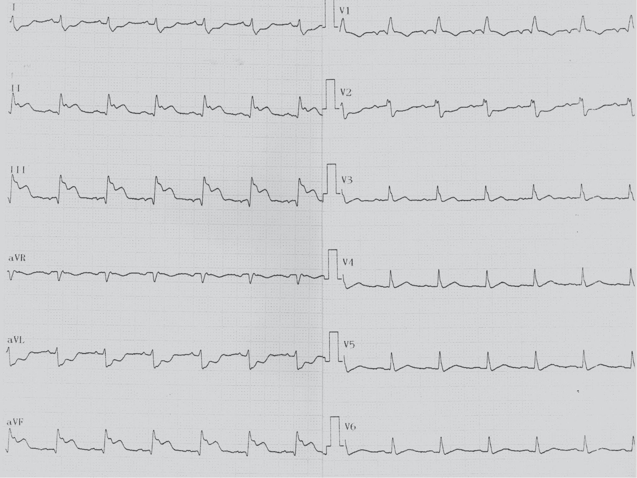
            Figure 1.

            ECG showed a sinus rhythm with a ST elevation and Q waves in inferior leads with specular ST-depression in leads V1, V2, I and aVL.

            Figure 2.

            Echographic images of the defect in parasternal view: on the left and lower portion of images, left ventricle is sectioned along its major axis and appear slightly hypertrophic. The right ventricle, in the upper and left portion of the images, appears dilated. (a) The entire course of the interventricular defect. (b) The defect in the projection that highlights its larger diameter. (c) Color-Doppler showing flux along the rupture. (d) Color-Doppler showing the left-to-right shunt in the right.

            Conclusion

            Early revascularization in AMI progressively reduced the prevalence of any complication of ischemic event, even more frequent like hypokinesis or arrhythmia. Although it is a rare complication of AMI, VSR is a life-threatening event and should be suspected in any patient with AMI, particular anterior and inferior/lateral ones, when a rapid hemodynamic deterioration is observed or when physical examination reveals a heart murmur or signs suggestive for right ventricle overload. Echocardiography is always needed and should be immediately performed in the ED. When VSR is diagnosed, a multidisciplinary team should collaborate to plan following management of patient, to obtain best outcome possible, according to high mortality related to this complication.

            What is new?

            The case report deals with a rare but severe complication of AMI, still associated with a high mortality rate. The peculiarity of our report is that the patient remained asymptomatic during observation in the emergency room and never had chest pain or signs of hemodynamic instability.

            Consent for publication

            A written consent was taken from the patient for publication.

            Ethical approval

            Ethical approval is not required at the institution to publish an anonymous case report.

            References

            1. Jones BM, Kapadia SR, Smedira NG, Robich M, Tuzcu EM, Menon V, et al.. Ventricular septal rupture complicating acute myocardial infarction: a contemporary review. Eur Heart J. 2014. Vol. 35(31):2060–8. https://doi.org/10.1093/eurheartj/ehu248

            2. Menon V, Webb JG, Hillis LD, Sleeper LA, Abboud R, Dzavik V, et al.. Outcome and profile of ventricular septal rupture with cardiogenic shock after myocardial infarction: a report from the SHOCK Trial Registry. Should we emergently revascularize Occluded Coronaries in cardiogenic shock? J Am Coll Cardiol. 2000. Vol. 36(Suppl. A):1110–6. https://doi.org/10.1016/S0735-1097(00)00878-0

            3. Birnbaum Y, Fishbein MC, Blanche C, Siegel RJ.. Ventricular septal rupture after acute myocardial infarction. N Engl J Med. 2002. Vol. 347:1426–32. https://doi.org/10.1056/NEJMra020228

            4. Moreyra AE, Huang MS, Wilson AC, Deng Y, Cosgrove NM, Kostis JB.. Trends in incidence and mortality rates of ventricular septal rupture during acute myocardial infarction. Am J Cardiol. 2010. Vol. 106:1095–100. https://doi.org/10.1016/j.amjcard.2010.06.013

            5. Arnaoutakis GJ, Zhao Y, George TJ, Sciortino CM, McCarthy PM, Conte JV.. Surgical repair of ventricular septal defect after myocardial infarction: outcomes from the Society of Thoracic Surgeons National Database. Ann Thorac Surg. 2012. Vol. 94:436–44. https://doi.org/10.1016/j.athoracsur.2012.04.020

            6. Thiele H, Kaulfersch C, Daehnert I, Schoenauer M, Eitel I, Borger M, et al.. Immediate primary transcatheter closure of postinfarction ventricular septal defects. Eur Heart J. 2009. Vol. 30:81–8. https://doi.org/10.1093/eurheartj/ehn524

            Summary of the case

            1 Patient (gender, age) Male, 74
            2 Final diagnosis Post-myocardial infarction VSR
            3 Symptoms None
            4 Medications
            5 Clinical procedure Reperfusion, Surgery
            6 Specialty Cardiothoracic Surgery

            Author and article information

            Journal
            European Journal of Medical Case Reports
            EJMCR
            Discover STM Publishing Ltd.
            2520-4998
            30 October 2020
            : 4
            : 10
            : 332-335
            Affiliations
            [1 ]Scuola di Specializzazione in Medicina di Emergenza-Urgenza, Università degli Studi di Milano Bicocca, Milano, Italy
            [2 ]U.O.C. di Medicina Generale, Emostasi e Trombosi, Fondazione IRCCS Ca' Granda Ospedale Maggiore Policlinico, Milano, Italy
            [3 ]Scuola di Specialzzazione in Medicina di Emergenza-Urgenza, Università degli studi di Milano, Milano, Italy
            Author notes
            [* ] Corresponding to: Matilde Conti Scuola di Specializzazione in Medicina di Emergenza-Urgenza, Università degli Studi di Milano Bicocca, Milano, Italy. Email: matilde_conti@ 123456yahoo.it
            Article
            ejmcr-4-332
            10.24911/ejmcr/173-1571084941
            7f98ea3e-f4a2-4153-9b1f-cea9d6bc4244
            © Matilde Conti, Enrico Rino Bregani, Mariairene Benedusi

            This is an open access article distributed in accordance with the Creative Commons Attribution (CC BY 4.0) license: https://creativecommons.org/licenses/by/4.0/) which permits any use, Share — copy and redistribute the material in any medium or format, Adapt — remix, transform, and build upon the material for any purpose, as long as the authors and the original source are properly cited.

            History
            : 15 March 2020
            : 18 May 2020
            Categories
            CASE REPORT

            cardiothoracic surgery,emergency PCI,echocardiography,case report,Acute myocardial infarction,post-ischemic ventricular rupture,heart murmur

            Comments

            Comment on this article