Background
Osteomyelitis in a younger age group is often seen as a common bone pathology due to multiple routes of infections. The entity may not be diagnosed in case of subtle findings or being at the subclinical level. This requires a thorough history and investigations for the complete diagnosis.
Case Report
A 14-year-old boy sustained right thigh injury while playing village sports game about 12 days back. There was no problem in the beginning, and he was given conservative treatment. The swelling appeared subsequently in the right thigh along with slight pain and restricted movements (Figure 1). There was a progressive increase in the swelling, and he reported to the hospital for further treatment. On local examination, there appeared an increase in the girth of the right thigh as compared to the contralateral side. Swelling was soft with fluctuations and measured 5.0 cm × 3.2 cm, and there was no increase in local temperature. Blood chemistry showed white blood cell count as 13,500/μL, erythrocyte sedimentation rate (ESR) as 40 mm in 1st hour, and C-elevated reactive protein (30 mgm/L). A systemic examination was unremarkable.
There was an increase in circumference (red star) of the right leg (white arrow) as compared to the left (green star).
Plain X-ray of the right thigh including the knee region was performed. No bony pathology was seen (Figure 2).
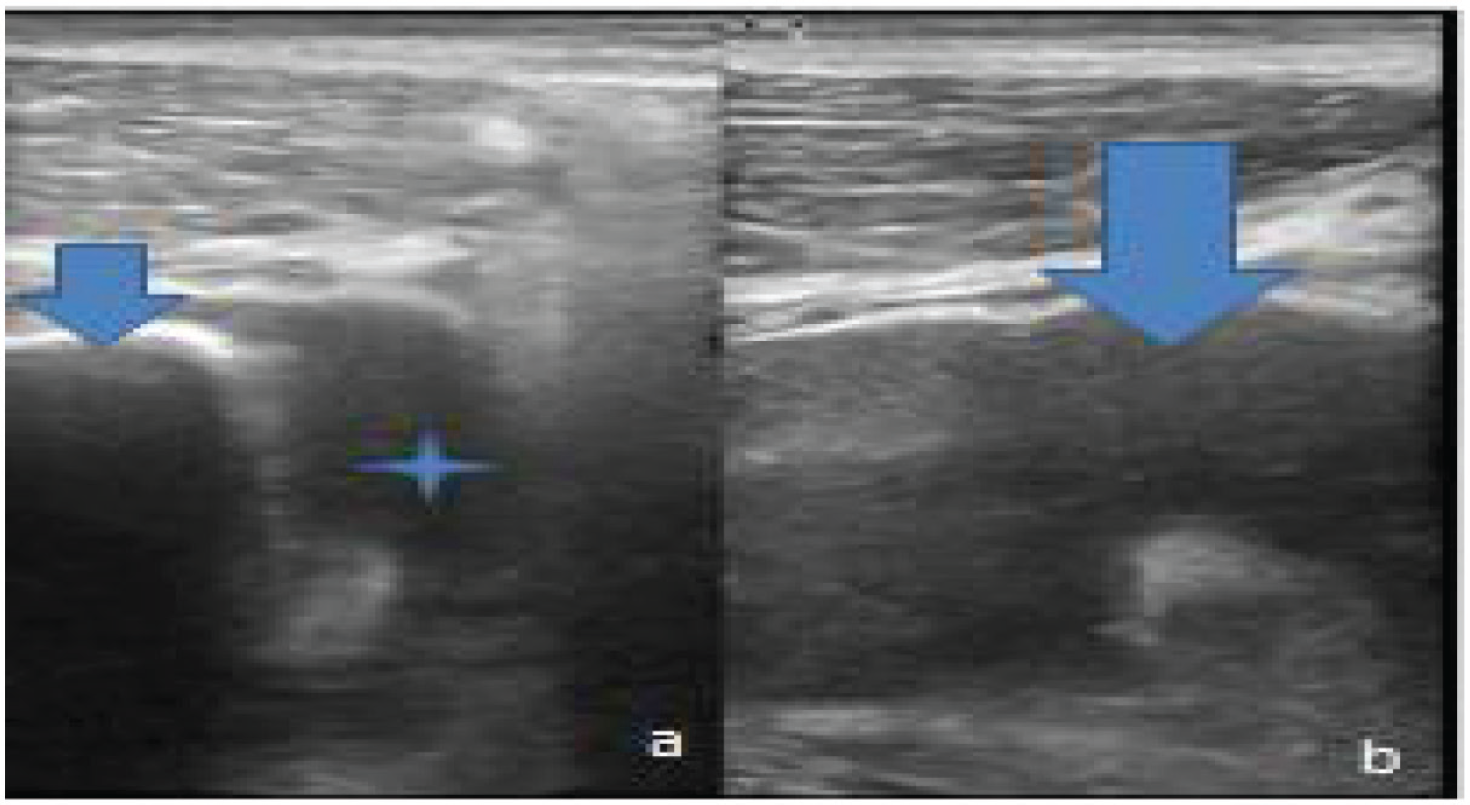
Ultrasonography of the right thigh. (a) Axial section shows the anechoic collection (blue star) with the adjoining femoral shaft as echogenic line (inverted blue arrow). (b) Long axis of the same region shows anechoic collection with some sediments within it (inverted blue arrow).
There was no evidence of underlying post-traumatic bony pathology. The patient was subjected to ultrasonography of the right thigh. There was an anechoic collection on the posteromedial aspect of the femur extending superoinferiorly. This collection was separated from the prepatellar collection (Figure 3a and b).
On color flow imaging, the region was without any abnormal blood flow (Figure 4a and b).
Computed tomography (CT) scanning was done; there was no evidence of fracture. However, some subtle low-density region in the posterior compartment was noticed in the mid-thigh region (Figures 5a–c and 6).
The patient was then subjected to magnetic resonance imaging (MRI) for further characterization (Figures 7- 9).
The working diagnosis of acute osteomyelitis with subperiosteal collection based on blood chemistry and radiological evaluation was considered. The aspirated fluid during surgical procedure was straw color. The cytological evaluation was performed using periodic acid–Schiff stain. The culture confirmed that the diagnosis of acute osteomyelitis showed the growth of Staphylococcus aureus. The patient was treated with a course of broad-spectrum antibiotic therapy with amikacin. A repeat sonographic examination showed complete resolution. The patient was discharged after 1 week of hospitalization and called for follow-up after a fortnight.
Discussion
There is a wide spectrum of differential diagnosis of the swelling of the thigh. Diagnostic imaging plays a vital role in diagnosis and further evaluation. Plain radiography of the region fits into a normal protocol followed by sonography. The fluid collection, periosteal involvement, and soft tissue abnormalities can easily be picked up on sonography with a high-frequency linear probe. CT can be an adjunct for the evaluation of bony elements. MRI study is excellent for the soft tissue involvement.
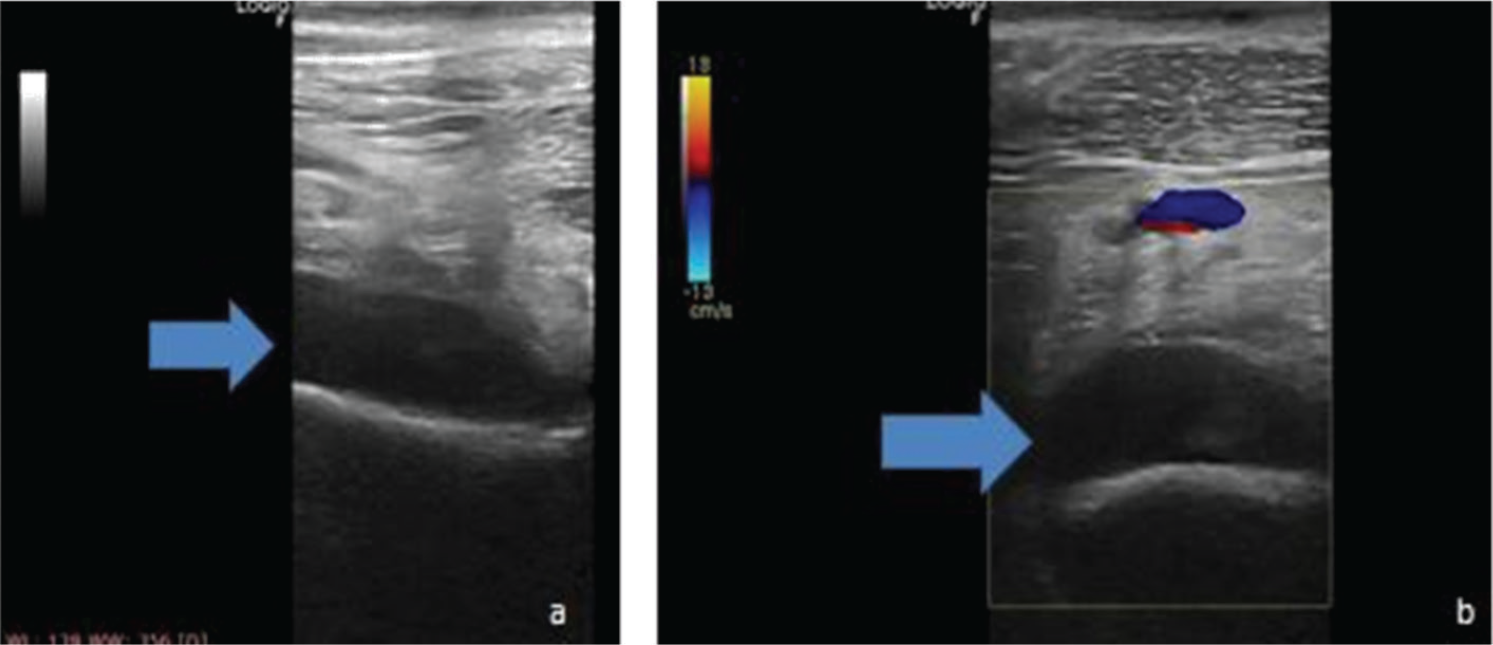
Color flow imaging. (a) Grayscale image at the collection region shows anechoic collection (blue horizontal arrow). (b) Switching on the color mode does not show any color within this collection (blue horizontal arrow) with minimal increase in vascularity in the surrounding region.
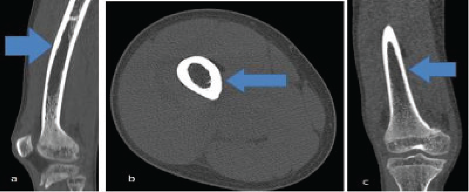
Non contrast-computerized tomography of the right thigh. 3D Multiplanar Reformation (MPR) image. (a) Sagittal projection does not show any abnormality in the thigh bone (blue horizontal arrow). (b) Axial section at mid femoral region shows well-maintained contour of the bone (blue horizontal arrow).(c) Coronal Section of right femur highlighting the normal bone (blue horizontal arrow).
The important clinic radiological features requiring thorough considerations in such cases are cortical tunneling, soft tissue swelling, focal cancellous lysis, and periosteal reaction.
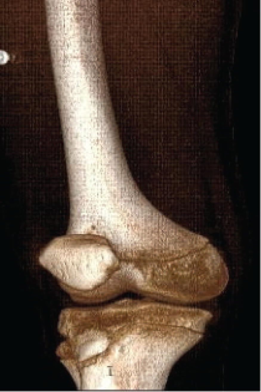
The right femur with the corresponding knee joint shows normal architecture in volumetric acquisition image. There is no abnormality or cortical break in the thigh bone.
Acute osteomyelitis may present with subperiosteal collection as was seen in our case. This infective process may be hidden for a long time before the diagnosis is made. Morel-Lavallee Lesion (MLL) is another post-traumatic entity which presents with hemolymph and necrotic fat collection. These are followed by injuries that are deep to the subcutaneous tissue and result due to the disruption of lymphatics and capillaries connecting them [1]. The swelling of the region becomes massive as the fluid with necrotic fat keeps on accumulating. This can lead to extensive focal necrosis if not treated promptly and adequately. This serosanguinous fluid, blood, and necrotic fat collection is present between the skin & subcutaneous tissue on one side and fascia on the other side [2]. Mellado and Bencardino classified these lesions into six types based on shape, MR signal intensity, enhancement pattern, and about the capsule [3]. These lesions should be differentiated from post-traumatic fat necrosis and coagulopathy hematomas. Although ultrasonography is the first line of investigation, MRI remains the gold standard for these types of lesions. Effusion gives characteristic findings of fluid attenuation interspersed with fat lobules. Management is conservative in type I and type II, whereas it is interventional or surgical in type III. Recurrence is common, and regular follow-up is required in these cases [4].

MRI images. (a) T1W axial section shows normal cortical hypointensity (white arrow) with central marrow hyperintensity. (b) T1W coronal section shows the normal interface between the femur and vastus muscle complex (red arrow). (c) Short tau inversion recovery (STIR) coronal image shows the clear interface between muscle and bone (green arrow).
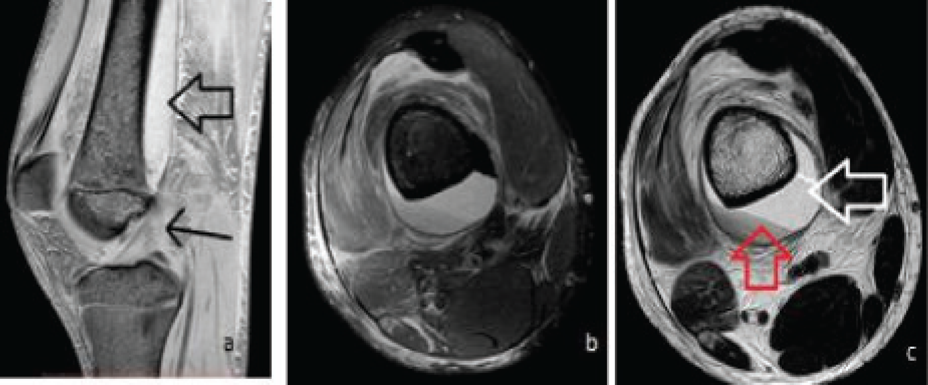
MRI contd. (a) Fast field echo image shows normal anterior cruciate ligament (thin black arrow) with hyperintense collection (wide horizontal arrow). (b) Proton density spectral presaturation inversion recovery axial section shows the hyperintense collection with sedimentation. (c) T2W axial section shows collection as hyperintensity (horizontal white arrow) with hypointense layering phenomenon of sedimentation (vertical red arrow).
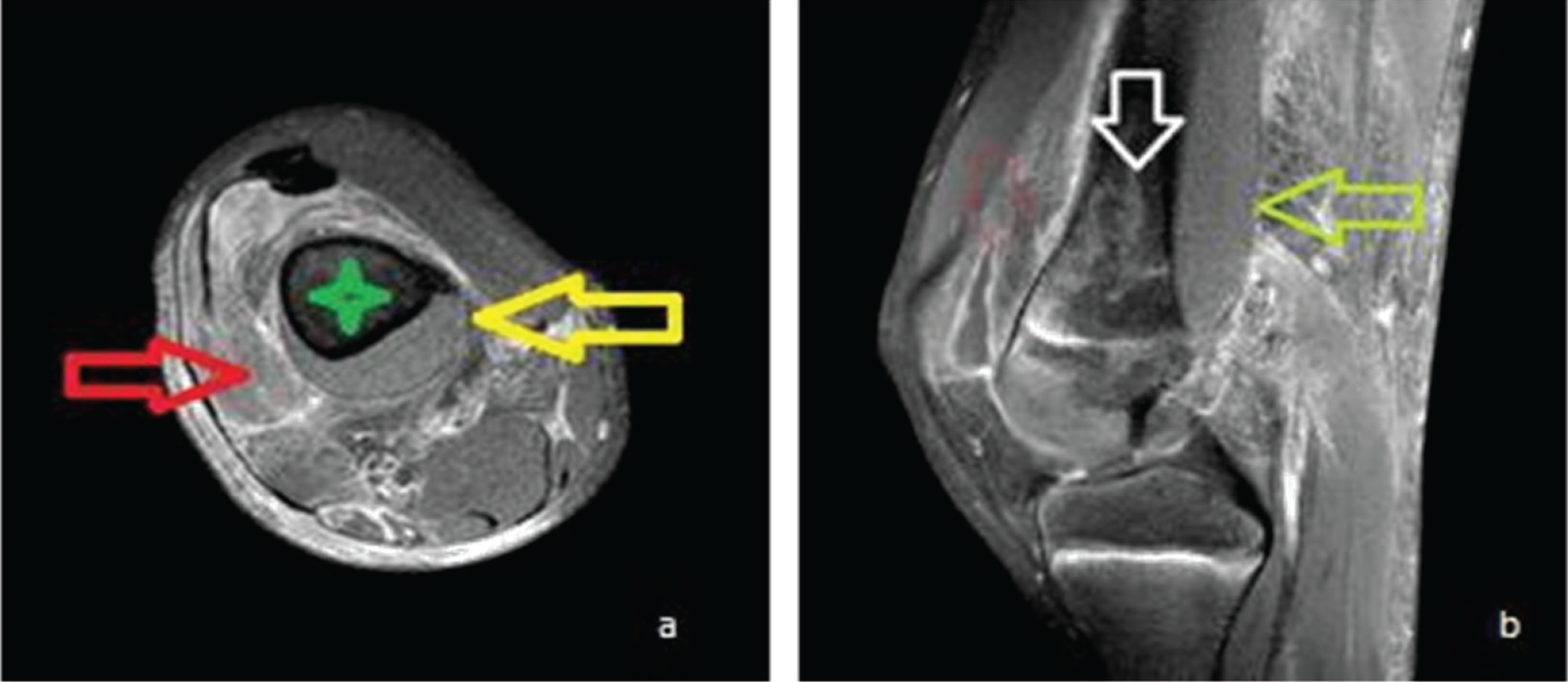
Contrast MRI. (a) T1W with fat saturation axial section shows well-defined fluid collection (yellow arrow) adjoining the femur (green star) with patchy enhancement of the surrounding muscles (red arrow). (b) Sagittal section of the same shows superoinferior extent of the collection (green arrow) with ill-defined femoral medullary intensity (white vertical arrow).
The knee is the most affected part in football players as per a study carried out by Tejwani et al. [5]. MR findings show classical findings of “fluid-fluid” level. Fat lobules in the lesions are best visualized by MRI because of high resolution and tissue characterization [6]. The “penumbra” sign is diagnostic in T1W study where there is peripheral portion showing a slight higher intensity as compared to cavity signal [7]. Cottias et al. [8] had presented one series of 21 osteomyelitis cases, which mimics bone tumors. Leukocytosis and raised ESR was seen in 9% and 30% of cases, respectively. Imaging can play a great role in the clinical and surgical management [9]. Nuclear medicine imaging is useful in detecting infection in 10–12 days’ period, but specificity is quite low [10]. An importance of diagnosis lies in early detection and management to avoid the complications [11].
Conclusion
Swelling of the thigh requires a thorough clinical and radiological evaluation. In the present case, swelling appeared after a trivial trauma without much complaint but was diagnosed as a case of acute osteomyelitis with subperiosteal collection. Cross--sectional imaging played a pivotal role in clinching the diagnosis as plain radiography was unremarkable. The cytological evaluation confirmed the diagnosis. The patient fully recovered after the surgical drainage and antibiotic therapy.


