44
views
0
recommends
+1 Recommend
1 collections
    0
    shares
      scite_
      0
      0
      0
      0
      Smart Citations
      0
      0
      0
      0
      Citing PublicationsSupportingMentioningContrasting
      View Citations

      See how this article has been cited at scite.ai

      scite shows how a scientific paper has been cited by providing the context of the citation, a classification describing whether it supports, mentions, or contrasts the cited claim, and a label indicating in which section the citation was made.

       
      • Record: found
      • Abstract: found
      • Article: found
      Is Open Access

      Pneumonia reveals a huge pleuropericardial cyst: a case presentation

      Published
      case-report
      Bookmark

            Abstract

            Background:

            Pleuropericardial cysts are rare and usually clinically silent, but can occasionally cause life-threatening complications. The majority of them is congenital due to developmental deficits and is most commonly found incidentally via routine radiography between the third and fifth decade of life.

            Case Presentation:

            A 37-year-old Caucasian woman who referred to the Emergency Room because of asthma exacerbation diagnosed suffering of pneumonia. During the follow-up and after antibiotics, chest X-Ray and computed tomography revealed a probably acquired large pleuropericardial cyst of unknown cause.

            Conclusion:

            We report an unusual case of possible infection which revealed an asymptomatic large pleuropericardial cyst which was detected accidentally via X-Ray and was left untreated due to its benign course.

            Main article text

            Background

            Mediastinal cysts are benign lesions and according to Takeda et al. represent 12%–18% or according to Gursoy et al. 20%–32% of all mediastinal masses [1,2]. Although cystic lesions surrounding the heart had been reported since 1854, according to the review of Lillie et al. [1], the first published pathoanatomic case series including 1 cyst was completed in 1903 [3].

            Pleuropericardial cysts (PPCs) account for 5%–10% of mediastinal tumors and 11% or 30% of mediastinal cysts. They are usually congenital [4,5]. However, other causes have been reported, including inflammatory, traumatic, and other causes [69]. Even though all ages may be affected, PPCs are most often detected between the third and fifth decade of life. The most common site of occurrence is the right cardiophrenic angle (51%–75%), followed by the left (28%–38%). PPCs usually have a diameter 3–15 cm and weigh 100–200 g. Most of PPCs are asymptomatic (50%–75%), so they are found post-mortem or incidentally on routine chest X-ray (CXR). When they are symptomatic, the symptoms are generally dominated by respiratory signs, such as chest discomfort or dyspnea, circulatory signs, and signs of nervous compression. Moreover, although they have almost always-benign clinical course, they can occasionally lead to complications due to compression of adjacent organs or inflammatory or other complications. As far as diagnosis is concerned, after the initial detection, further imaging, including multi detector computed tomography, Magnet Resonance Imaging, and other methods are used complementarily to confirm the diagnosis mainly in difficult cases. Given that PPCs are usually asymptomatic, and it has not been reported to have malignant potential, the majority of them can be left without treatment. When management is required, surgical resection by means of traditional open surgery or minimally invasive methods is considered to be the gold standard and along with percutaneous aspiration are the methods that have been mostly performed [10].

            We report below a peculiar case of a 37-year-old woman who referred to the Emergency Room (ER) because of suspected asthma exacerbation and computed tomography (CT) revealed a probably acquired large PPC of unknown cause.

            Case Presentation

            A 37-year-old Caucasian woman with medical history of focal epilepsy and asthma, referred to our ER because of suspected asthma exacerbation (palpitations and dyspnea). Clinical investigation was suspected of pneumonia. Biochemical tests revealed a slight increase of C-reactive protein and leucocytosis which raised the suspicion of infection. A CXR revealed an intermediate pneumonia (Figure 1a). A control radiograph 2 weeks later under Penicillin treatment revealed remaining findings, but the patient was asymptomatic (Figure 1b). A new CXR 2 months after the first demonstrated a slight progress of chest mass (Figure 1c). A contrast thoracic CT (Siemens SOMATOM Sensation 64 slice) with a 3 mm slice thickness, confirmed the presence of an anterior basal right-sided PPC sized 5.5 cm craniocaudally, 5 cm anteroposterior, and 6 cm laterally (Figure 2) without contrast agent assuming, pathologically enlarged lymph nodes and pleural effusion.
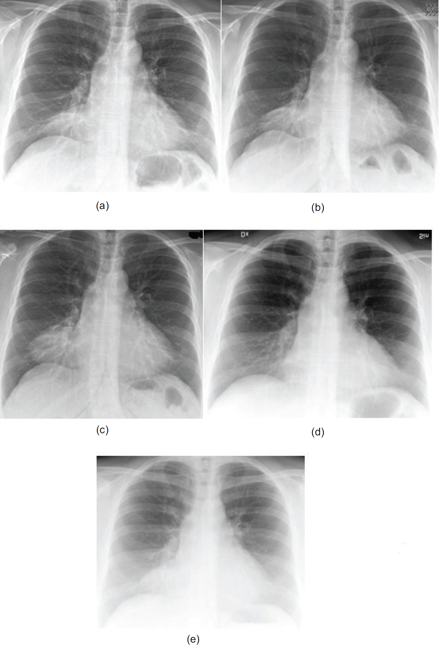
            Figure 1.

            (a) First CXR. Intermediate pneumonia. (b) Control CXR 2 weeks after the first. Remaining findings while patient was asymptomatic under penicillin treatment. (c) Control CXR 2 months after the first. Slight progress of chest mass while patient was still asymptomatic. (d) Previous CXR in the last 8 years. (e) Previous CXR in the last 8 years.

            According to the literature, on CT scan, the PPC is thin-walled, well-marginated, oval homogeneous mass, usually unilocular, while multilocular cysts have also been reported. Their attenuation is low (0–20 HU) although sometimes it may be a little higher than water density (30–40 HU). This is probably interpreted by a high protein and cells content due to bleeding or infection [10]. As they are commonly avascular, they don’t enhance with contrast agents. Abdominal CT and echocardiogram were performed and both were normal without pathological focal changes. The differential diagnostic approach was challenging. Radiological investigation combined with a normal laboratory control excluded pericardial or pleural effusion, aneurysms, fluid-filled superior aortic recess, varicose pericardial vein, lipoma, hydatic heart disease, germ cell tumors, diaphragmatic hump, mecechymal tumors, brochogenic carcinoma, and mediastinal abscess, as a possible diagnosis. After the examination of previous radiographies in the last 8 years, this cyst was not observed (Figure 1d and e). Therefore, this event constitutes a peculiar case of an asymptomatic probably acquired PPC with unknown cause according to patient's medical history. After consultation with lung specialists and thoracic surgeons, ultrasound-guided punctuation was recommended in the event of symptoms. Video-assisted thoracoscopic surgery could be a further choice of management in case of cyst's refill within a short period after punctuation. Eventually, no surgical intervention was performed, as the patient did not develop any cyst-associated symptoms, after a 5-year follow up.

            Figure 2.

            A contrast thoracic CT confirmed the presence of an anterior basal right-sided PPC (inside the yellow circle) sized 5.5 cm craniocaudally, 5 cm anteroposterior and 6 cm laterally without pathologically enlarged lymph nodes and pleural effusion.

            Conclusion

            PPCs are rare and usually clinically silent, but can occasionally cause life-threatening complications. The majority of them is congenital due to developmental deficits and is most commonly found incidentally via routine radiography between the third and fifth decade of life. The management of a PPC should be based on an algorithm taking into consideration cyst's size, shape, and compressibility as well as clinical presentation and patient’s fitness and preference. Surgical resection by means of traditional open surgery or minimally invasive methods is considered to be the gold standard and along with percutaneous aspiration are the methods that have been mostly performed. Finally, we reported an unusual case of possible infection which revealed an asymptomatic large PPC which was detected accidentally via CXR and was left untreated. Not having observed in earlier imaging studies this cyst is believed to be acquired, but the exact etiology has remained unknown as aspiration which could exclude inflammatory causes has not been required.

            What is new?

            To our knowledge, it is the first time that a pleuropericardial cyst reveals in an asymptomatic patient who is presented to emergency department with pneumonia. Patients’ infection-related symptoms disappeared after antibiotics, but the radiological investigation showed an unchanged huge pleuropericardial cyst.

            List of Abbreviations

            CT

            Computed tomography

            CXR

            Chest X-ray

            ER

            Emergency room

            PPCs

            pleuropericardial cysts

            Consent for publication

            Written informed consent for the paper to be published (including images/videos) was obtained from the patient. She understood that the information should be published without her name attached, but that full anonymity could not be guaranteed. She understood that the text and any pictures or videos published in the article would be freely available on the internet and may be seen by the general public. The pictures, videos and text may also appear on other websites or in print, may be translated into other languages or used for commercial purposes. She had been offered the opportunity to read the manuscript.

            Ethical approval

            Ethical approval is not required in our institution for publishing an anonymous case report.

            References

            1. Lillie WI, Mc DJ, Clagett OT. Pericardial celomic cysts and pericardial diverticula; a concept of etiology and report of cases. J Thorac Surg. 1950. Vol. 20(3):494–504

            2. Takeda S, Miyoshi S, Minami M, Ohta M, Masaoka A, Matsuda H. Clinical spectrum of mediastinal cysts. Chest. 2003. Vol. 124(1):125–32. [Cross Ref]

            3. Rohan A. Ueber divertikel-und cystenbildung am perikard [in German]. Prag Med Wochschr. 1903. Vol. 28:461–4

            4. Lambert AVS. Etiology of thin-walled thoracic cysts. J Thorac Surg. 1940. Vol. 10(1):1–7

            5. Murray JFaN JA. Textbook of respiratory medicine. Philadelphia, PA: W.B. Saunders Company. 1994. 2146–7

            6. Maisch B, Seferovic PM, Ristic AD, Erbel R, Rienmuller R, Adler Y, et al.. Guidelines on the diagnosis and management of pericardial diseases executive summary; The Task force on the diagnosis and management of pericar-dial diseases of the European society of cardiology. Eur Heart J. 2004. Vol. 25(7):587–610. [Cross Ref]

            7. Patel J, Park C, Michaels J, Rosen S, Kort S. Pericardial cyst: case reports and a literature review. Echocardiography. 2004. Vol. 21(3):269–72. [Cross Ref]

            8. Pugliatti P, Donato R, Crea P, Zito C, Patane S. Image diagnosis: pericardial cyst in a dialysis patient. J Cardiovasc Ultrasound. 2016. Vol. 24(2):177–8. [Cross Ref]

            9. Saldana Duenas C, Hernandez Galan A. Posttraumatic pericardial cyst. An Sist Sanit Navar. 2015. Vol. 38(3):475–8. [Cross Ref]

            10. Koumantzia C SN, Eleftheriou A. Pleuropericardial cyst: a review of the literature. J Cardiol Cardiovasc Sci. 2019. Vol. 3(4):1–29. [Cross Ref]

            Summary of the case

            1 Patient (gender, age) 37-year-old Caucasian woman
            2 Final diagnosis Pleuropericardial cyst
            3 Symptoms Asthma exacerbation, palpitations, dyspnea
            4 Medications antibiotics
            5 Clinical procedure Clinical investigation, X-Rays, thoracic CT, radiological and clinical follow-up
            6 Specialty Cardiology

            Author and article information

            Journal
            European Journal of Medical Case Reports
            EJMCR
            Discover STM Publishing Ltd.
            2520-4998
            30 March 2020
            : 4
            : 3
            : 93-96
            Affiliations
            [1. ]Department of Neurology and Department of Biomedical and Clinical Sciences, Division of Neurobiology, Linköping University, Linköping, Sweden
            [2. ]Primary Health Care Center Tannefors, and Department of Medical and Health Sciences, Linköping University, Linköping, Sweden
            Author notes
            [* ] Correspondence to: Andreas Eleftheriou Department of Neurology and Department of Biomedical and Clinical Sciences, Division of Neurobiology, Linköping University, Linköping, Sweden. andelef2002@ 123456yahoo.gr ; andreas.eleftheriou@ 123456regionostergotland.se
            Article
            ejmcr-4-93
            10.24911/ejmcr/173-1549190787
            b0bbb8c0-88d3-4ce1-a164-ac3e4a1248e3
            © Andreas Eleftheriou, Nikolaos Saridakis, Christina Koumantzia

            This is an open access article distributed in accordance with the Creative Commons Attribution (CC BY 4.0) license: https://creativecommons.org/licenses/by/4.0/) which permits any use, Share — copy and redistribute the material in any medium or format, Adapt — remix, transform, and build upon the material for any purpose, as long as the authors and the original source are properly cited.

            History
            : 05 February 2019
            : 24 March 2020
            Categories
            CASE REPORT

            pneumonia,asthma exacerbation,mediastinum,Pleuropericardial cyst

            Comments

            Comment on this article