49
views
0
recommends
+1 Recommend
1 collections
    0
    shares
      scite_
      0
      0
      0
      0
      Smart Citations
      0
      0
      0
      0
      Citing PublicationsSupportingMentioningContrasting
      View Citations

      See how this article has been cited at scite.ai

      scite shows how a scientific paper has been cited by providing the context of the citation, a classification describing whether it supports, mentions, or contrasts the cited claim, and a label indicating in which section the citation was made.

       
      • Record: found
      • Abstract: found
      • Article: found
      Is Open Access

      Atypical Osteoid Osteoma of sacral region in pediatric age group: a case report

      Published
      case-report
      Bookmark

            Abstract

            Background:

            Osteoid osteomas are benign lesions of osteoblastic activity. These are frequently found in many instances where there is pain of unexplainable background. But many a times the entity is found as incidental finding while the patient is being evaluated for some other reason.

            Case presentation:

            We present 15 years old boy who reported for backache of two months duration. This used to get worsened at night time and with some relief with non-steroid anti-inflammatory drugs (NSAID). Plain X-ray did not show any abnormality. The child underwent magnetic resonance imaging (MRI) which has given some clue for further evaluation by non-contrast computerized tomography (NCCT) of lumbosacral region. This had brought out the diagnosis which was subsequently confirmed by radio nuclear studies.

            Conclusion:

            Pediatric age group backache is rare phenomenon and thorough search is required to investigate the cause. Various imaging modalities often locate the pathology if evaluated properly. This helps in the management of the case.

            Main article text

            Background

            Jaffe put osteoid osteoma lesion in a separate category in 1935 [1]. This is constituted by central nidus rich in osteoid tissue with variable amount of calcification. There is normal sclerotic bone tissue in the surrounding region. This can either be at solitary or multiple locations in different bones.

            Case presentation

            15-years old boy reported to the pediatric outpatient department with complaints of backache since two months duration. This pain was severe at night and used to get relieved with analgesics. Then the intensity of pain increased and the patient reported to the hospital accordingly. There was no history of trauma or any other systemic disease. He was of averagely built physique. All the biochemical parameters were within normal limits. Systemic examination was unremarkable. Plain lumbo-sacral and abdomen skiagram were normal (Figure 1).

            The boy was subjected to cross-sectional imaging. Magnetic resonance imaging study was performed which revealed subtle signal intensity changes in sacral region in T1W, T2W and STIR images (Figure 2 and 3). There was post gadolinium enhancement of the lesion (Figure 4).

            NCCT of lumbosacral region was performed with the consent of the patient. This revealed a radiolucent focus with central radiodense nidus which measured 0.9 × 0.9 cm (Figure 5a, b and c). The working diagnosis of osteoid osteoma was kept in consideration. Later, the patient underwent radio-nuclear study which confirmed the diagnosis. This had shown increased uptake of the lesion region (Figure 6a and b). The child had been put on symptomatic treatment with future plan for the radiofrequency ablation. Three months follow-up had been advised before deciding about any RF ablation or surgical intervention.
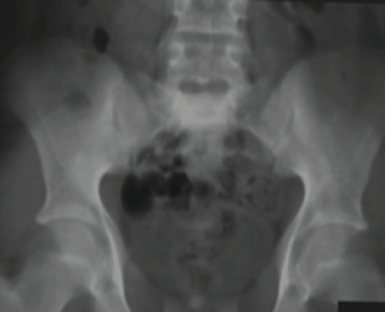
            Figure 1:

            Plain pelvis skiagram including sacral region antero-posterior view. There is no evidence of any lesion or pathology seen in the sacral region.
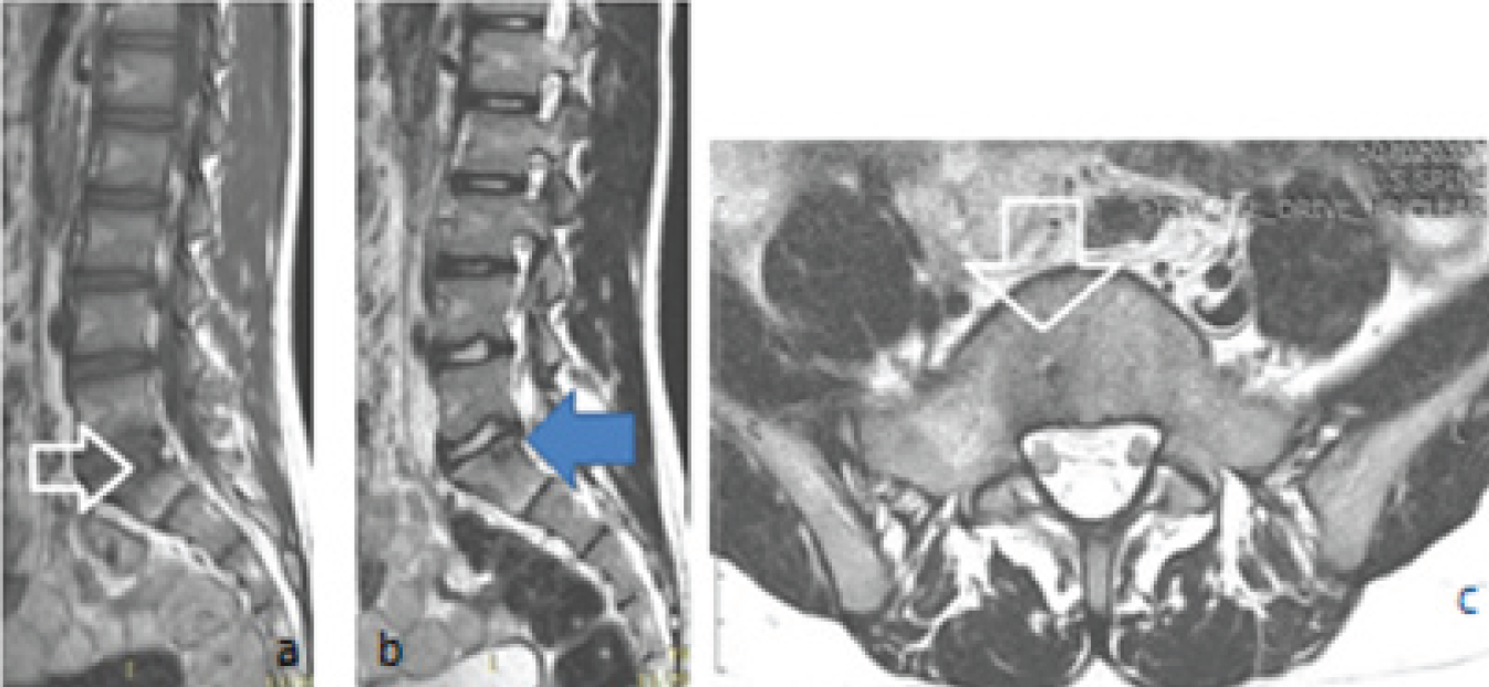
            Figure 2:

            Magnetic resonance images, (a) T1W sagittal section shows a slightly low intensity region in the body of S1 sacral vertebra (white hollow arrow), (b) T2W sagittal section reveals mixed intensity lesion at the same location (blue horizontal arrow), (c) T2W axial section shows round hypointense lesion with surrounding hyperintensity (white hollow arrow).
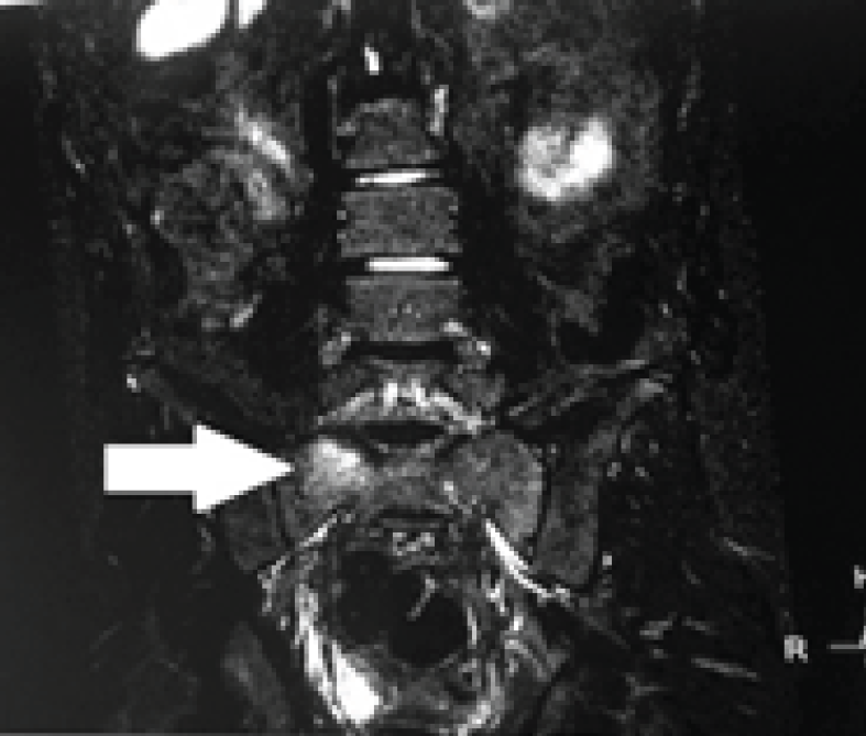
            Figure 3:

            STIR magnetic resonance image show subtle hyperintensity in the body of S1 sacral vertebra suggestive of edema (solid white arrow).
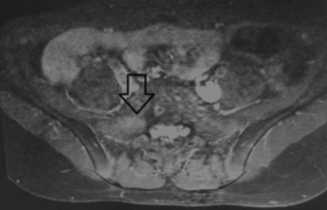
            Figure 4:

            Contrast enhanced T1W fat saturated image. There is enhancement seen on the right side of the body of S1 vertebra (black vertical hollow arrow). The region corresponds to the location of the osteoid osteoma.

            Figure 5:

            NCCT of pelvis and sacral region, (a) axial section shows small hypodense lesion with hyperdense central nidus (blue vertical arrow), (b) Coronal reformatted image shows the same lesion in the body of S1 vertebra (black hollow vertical arrow), (c) Sagittal reformatted image shows S1 vertebral hyper density (vertical hollow black arrow).
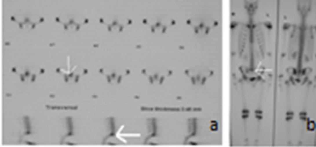
            Figure 6:

            Nuclear scan for the bones, (a) axial sections (vertical white arrow) and sagittal section (horizontal white arrow) shows the increased early uptake of the tracer in the S1 vertebral lesion, (b) whole body scan shows the increased abnormal uptake at the sacral region (white horizontal arrow).

            Discussion

            Osteoid osteomas are benign bony lesions with osteoblastic activity. Though these can occur in any age group but 10-30 years is the most common group. Barei et al have noticed that the majority of osteoid osteomas occur below 20 years of age [2]. Lower extremities are the most common for their occurrence. Femur is the most common bone affected and cortical region of the long bones is the common most site.80-90% cases occur in the shafts of long bones and rest in other different locations. 7-20% have been observed in the spinal region. 78% of the cases in the lumbar region have been associated with scoliosis [3,4]. Following is the predilection for the occurrence in the spine as per the region involved:

            • 59% Lumbar spine

            • 27% Cervical region

            • 12% Thoracic region

            • 2% Sacral region

            Well at al have shown that in the majority of the cases there is a predilection for the posterior elements and the vertebral body is rarely involved [5]. Male are affected more than females and ratio is 2-3:1. Our case was male and falls in the younger age group. He had shown the involvement of the first sacral vertebral body which is slightly unusual site. The entity requires to be differentiated from eosinophilic granulomas, Brodie abscess and other cystic bone pathologies. Radiographically, these lesions may not be picked up at the first time because of their atypical complaints and presentations. Plain x-ray and NCCT scan delineate the central nidus with surrounding sclerotic zone. Bone scan confirms the diagnosis by early radiotracer uptake and delayed accumulation. This differentiates it from many other similar types of pathologies. Atar et al have divided this entity into two stages as acutely painful which is early and recovery stage which is later [6]. The second stage, in fact, corresponds to the healing because of ossification of the nidus. The management of these lesions in childhood is a challenge for the clinicians. There are various treatment options for the entity and radiofrequency ablation remains the choice in selected cases with the appropriate setting [7]. The lesion should not be near to the thecal sac and there should be enough cortical bone in the surrounding region. Air in the region can be of benefit being non-conduction of heat.

            Conclusion

            Backache in children is slightly uncommon feature. When the child is investigated for unexplained backache than the possibility for the search of other differentials must be kept in mind. There can be atypical presentation of osteoid osteomas at some uncommon sites. There may be a small navigating clue which can lead to the diagnosis. These are usually diagnosed through cross sectional imaging like MRI and NCCT. The management depends on the location, size and the symptoms of the patient.

            Acknowledgements

            We are thankful to Mr. Nitish Vermani, Rajdeep and Raushan Singh from the Faculty of Allied Health Sciences SGT University for carrying out general radiography and acquiring the MRI and NCCT images for this study.

            List of Abbreviations

            LS

            Lumbosacral Spine

            NCCT

            Non Contrast Computerized Tomography

            RF

            Radiofrequency

            STIR

            Short Tau Inversion Recovery

            Conflict of Interests

            None

            Funding

            None

            Consent for publication

            Informed consent was obtained from the patient to present and publish this case.

            Ethical approval

            Ethical approval is not required at our institution for publishing a case report in a medical journal.

            Authors’ contribution

            All authors contributed in the management of the patient. All of them have also contributed to the drafting, revising and final editing of the manuscript.

            References:

            1. Atar D, Lehman WB, Grant AD.. Tips of the trade. Computerized tomography--guided excision of osteoid osteoma. Orthop Rev. 1992. Vol. 21:1457–8

            2. Barei DP, Moreau G, Scarborough MT.. Percutaneous radiofrequency thermalablation of osteoid osteoma. Operative Techniques in Orthopaedics. n.d.. Vol. 9:72–8. [10.1016/S1048-6666(99)80024-X]

            3. Burn SC, Ansorge O, Zeller R, Drake JM.. Management of osteoblastoma and osteoid osteoma of the spine in childhood. J Neurosurg Pediatr. 2009. Vol. 4:434–8. [10.3171/2009.6. PEDS08450]

            4. JAFFE HL.. “osteoid-osteoma”: benign osteoblastic tumor composed of osteoid and atypical bone. Archives of Surgery. 1935. Vol. 31:709–28. [10.1001/archsurg.1935.01180170034003]

            5. Kan P, Schmidt MH.. Osteoid osteoma and osteoblastoma of the spine. Neurosurg Clin N Am. 2008. Vol. 19:65–70. [10.1016/j.nec.2007.09.003]

            6. Wells RG, Miller JH, Sty JR.. Scintigraphic patterns in osteoid osteoma and spondylolysis. Clin Nucl Med. 1987. Vol. 12:39–44

            7. Zileli M, Cagli S, Basdemir G, Ersahin Y.. Osteoid osteomas and osteoblastomas of the spine. Neurosurg Focus. 2003. Vol. 15:E5

            Summary of the case

            Patient (gender, age) 1Male, 15 year old
            Final Diagnosis 2Sacral osteoid osteoma
            Symptoms 3Low backache
            Medications (Generic) 4Conservative with three months follow up
            Clinical Procedure 5Contrast MRI
            Specialty 6Orthopedics, pediatrics
            Objective 7To find out the cause of backache
            Background 8Young boy with backache
            Case Report 9Cross-sectional imaging for backache in pediatric age group
            Conclusions 10NCCT and CE MRI of LS and Pelvis in diagnosis
            MeSH Keywords 11Osteoid osteoma, case report, backache, NSAID, MRI, NCCT

            Author and article information

            Journal
            European Journal of Medical Case Reports
            EJMCR
            Discover STM Publishing Ltd.
            2520-4998
            30 January 2017
            : 1
            : 1
            : 28-31
            Affiliations
            [1 ]Department of Pediatrics, SGT Medical College Budhera (Gurgaon), India.
            [2 ]Department of Radio-diagnosis, SGT Medical College Budhera (Gurgaon), India.
            [3 ]Faculty of Allied & Health Sciences, SGT University Gurgaon, Haryana, India.
            Author notes
            [* ] Correspondence to: Dr. Shashi Sharma, Department of Pediatrics, SGT Medical College, Budhera (Gurgaon), India. Email: shashi.s986@ 123456gmail.com
            Article
            ejmcr-1-28
            10.24911/ejmcr/1/7
            f5922026-bbd6-4967-bd48-865cf9046b27
            © Shashi Sharma, Sakshi Dewan, Naveen Bhardwaj, Mir Rizwan Aziz, Shilpa Singh, Bharat Bhushan Sharma

            This is an open access article distributed in accordance with the Creative Commons Attribution (CC BY 4.0) license: https://creativecommons.org/licenses/by/4.0/) which permits any use, Share — copy and redistribute the material in any medium or format, Adapt — remix, transform, and build upon the material for any purpose, as long as the authors and the original source are properly cited.

            History
            : 27 November 2016
            : 11 January 2017
            Categories
            CASE REPORT

            NSAID,backache,case report,NCCT,Osteoid osteoma,MRI

            Comments

            Comment on this article