Background
Pyramidal lobe is remnant of thyro-glossal duct and is rising upwards from right or left lobe or isthmus of thyroid gland. Prevalence of pyramidal lobe is reported between 15 to 75% [1]. It may have functional thyroid tissue (Usually low functioning than normal thyroid tissue). On the therapeutic floor, determination of pyramidal lobe pre-operatively is important in relation to Grave’s disease to avoid recurrence. Diagnosis beforehand is also important in the treatment of other diseases like thyroid cancer etc. It helps to plan proper treatment and avoid recurrence [2].
Radionuclide pertechnetate (99mTcO4) thyroid scintigraphy is widely used for evaluation of neck nodules whether functioning or non-functioning nodules. Hyper-metabolic nodules show intense radionuclide uptake with suppressed background activity. Hot nodules in thyroid gland are routinely diagnosed on 99mTcO4 thyroid scintigraphy. Ectopic thyroid tissue has been reported in literature mostly in head and neck region. Common sites are pyramidal lobe, lingual thyroid, trachea, submandibular, palatine tonsils and lateral cervical regions. Detection of pyramidal lobe by thyroid scintigraphy ranges from 4.2% to 40% [3]. We are reporting a case of hot nodule in pyramidal lobe as visualized on 99mTcO4 thyroid scintigraphy which is rare and not previously reported in literature.
Case presentation
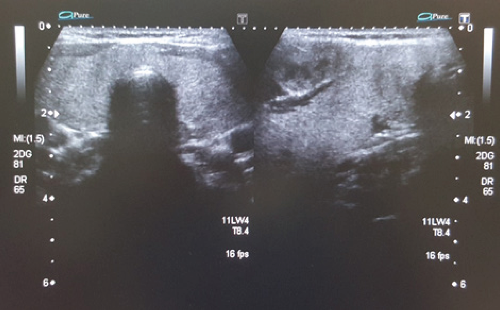
A 20 year old female presented in outdoor department with a palpable nodule in front of the neck adjacent to thyroid cartilage. Clinically she was euthyroid. Nodule was moving slightly with deglutition while no movement was noted on protrusion of tongue. Swelling seemed to be a thyroid nodule. Thyroid function tests (TFTs) confirmed euthyroid status. The patient was subjected to evaluation with neck ultrasound, which showed a well-defined 26mm nodule in the pyramidal lobe arising from the right half of isthmus (Figure: 1). Nodule showed central tiny areas of cystic degeneration with increased doppler flow in the mid region while significantly increased doppler flow at its periphery depicting hypermetabolic activity (Figure: 2). Her 99mTcO4 thyroid scan was performed with Infinia dual head gamma camera equipped with low energy high-resolution collimators at 140Kev peak with 20% energy window. Apart from radiotracer uptake in thyroid gland, scan showed an important finding of intense 99mTcO4 focal uptake adjacent to the medial border of right lobe of thyroid (Figure: 3-A). An additional image acquired, with a hot marker carefully placed on the palpable nodule, confirming the intense uptake to be in palpable nodule (Figure: 3-B). The final diagnosis was hot nodule in the pyramidal lobe arising from right half of isthmus. Later excisional biopsy of the nodule confirmed that it was thyroid tissue.

High resolution ultrasound images showing nodule in the pyramidal lobe arising from the right lobe of thyroid gland.
Discussion
Hot nodules are common in both lobes of thyroid. In pyramidal lobe, it is not seen before. It is a unique case and not yet reported in the literature.
The pyramidal lobe is related to the distal portion of the thyro-glossal duct [4]. The pyramidal lobe differs in shape, size and position. Its course is usually upwards, lying in midline or laterally depending on its attachment with thyroid gland which may be form superior border of the isthmus, medial or superior aspects of either of thyroid lobes [5]. In literature, pyramidal lobe was most frequently observed to be originated from left lobe as compared to right lobe and isthmus, with frequency of 15% to 75% as per anatomy books [6–8]. In cadaver studies, the prevalence of pyramidal lobe reported, ranges from 28.9% - 55% [1]. A study done on South Korean population using computed tomography has demonstrated the incidence of pyramidal lobe to be 44.6% [9]. The occurrence of pyramidal lobe shows some gender predominance though little data is available. Some articles suggest that it is more prevalent among females, while according to some authors it is more often detected among males [10,11].
Thyroid cells in the pyramidal lobe are usually hypo-functional; however, they can become active after excision of the functioning thyroid tissue/ablation of the thyroid tissue in hyperthyroid patients. In such cases, recurrent hyperthyroidism may develop especially in cases of total thyroidectomy due to Graves’ disease. The presence of thyroid cancer in pyramidal lobe has also been reported in the literature [2,12]. In literature, the rate of detection of pyramidal lobe by 99mTcO4 thyroid scintigraphy ranges from 4.2 to 40% [13]. This incidence of detection of pyramidal lobe is quite low when compared to anatomic and surgical methods. The attributed reason behind this is the thin structure of pyramidal lobe with practically hypo-functional thyroid cells [14].
In patients with carcinoma thyroid, residual pyramidal lobe may hamper the upsurge of TSH post operatively. This is really important in the patients who are planned to undergo postoperative radioactive Iodine-131 (RAI-131) treatment, therefore, affecting the effectiveness of RAI-131 treatment. This is why it is really important to know the presence and location of pyramidal lobe. These patients might need RAI-131 treatment after thyroid surgery. It is also very important to differentiate between mid-line esophageal activity and pyramidal lobe on the radionuclide scintigraphic images. Patients must drink a glass of plain water just before radionuclide imaging, to avoid possible esophageal artifact. Additional anterior oblique images may also serve the purpose as pyramidal tissue is located anteriorly while esophageal activity is located posteriorly to the thyroid gland [15]. Levy et al reported the incidence of pyramidal lobe to be 43% in diffuse toxic goiter / Graves’ disease, 11% in nodular goiters, 10% in solitary functioning nodules and in 17% patients with normal thyroid glands [14]. According to Zivic et.al, the incidence of pyramidal lobe was greater in the younger age group compared to older age. The reported reasoning behind this was high incidence of Graves’ disease in younger age group [15]. Pyramidal lobe can also be localized by radiological modalities such as high-resolution ultrasound (Usg), computed tomography (CT) or magnetic resonance imaging (MRI).
Before determining treatment options, proper diagnosis is necessary to confirm the nature of lump in almost mid line. Likely differential diagnosis will include lymph node, lipoma, thyro-glossal cyst and lump with thyroid tissue. Lump with thyroid tissue in mid line may be ectopic thyroid tissue, thyroidal tissue in thyro-glossal cyst, or nodule in the pyramidal lobe. The patient was evaluated with neck ultrasound, which showed a well-defined 26mm nodule in pyramidal lobe arising from the right half of isthmus. Radionuclide (99mTcO4) thyroid scintigraphy is one of the effective tools in diagnosis of pyramidal lobe and it pathologies. In our case, radionuclide (99mTcO4) thyroid scintigraphy localized an area of intense uptake in medial aspect of right lobe of thyroid gland. It was in a clinically palpable nodule. Area of intense uptake to be a nodule in the pyramidal lobe, collectively making preliminary diagnosis of hot nodule in pyramidal lobe arising from right half of isthmus. The presence of hot nodule in pyramidal lobe is very rare, as thyroid cells in the pyramidal lobe are hypo-functional i.e. practically non-functional. Diagnosis of hot nodule in pyramidal lobe is further difficult as it appears to be in any of the thyroid lobes on radionuclide (99mTcO4) thyroid scintigraphy. There is no difference in treatment protocol of hot nodule in pyramidal lobe or elsewhere in the thyroid gland. There are many different treatment options for hot nodule in pyramidal lobe e.g. just observe, RAI-131 ablation and surgery. In this particular case (hot nodule in pyramidal lobe with normal thyroid gland in situ) surgery is the choice of treatment. It was later removed to end the stigma of nodule in the neck. It proved to be a thyroid tissue. The patient maintained biochemically euthyroid status.


