87
views
0
recommends
+1 Recommend
1 collections
    0
    shares
      scite_
      0
      0
      0
      0
      Smart Citations
      0
      0
      0
      0
      Citing PublicationsSupportingMentioningContrasting
      View Citations

      See how this article has been cited at scite.ai

      scite shows how a scientific paper has been cited by providing the context of the citation, a classification describing whether it supports, mentions, or contrasts the cited claim, and a label indicating in which section the citation was made.

       
      • Record: found
      • Abstract: found
      • Article: found
      Is Open Access

      Ischio-pubic osteochondritis (Van Neck-Odelberg disease): A case report

      Published
      case-report
      Bookmark

            Abstract

            Background:

            Ischio-pubic Osteochondritis is characterized by an atypical ossification pattern of the ischio-pubic synchondrosis. Radiologically it may mimic stress fracture, neoplasm, osteomyelitis, or post-traumatic osteolysis. 99mTc-MDP bone scintigraphy with SPECT/CT is one of the effective tools for its diagnosis.

            Case presentation:

            A male patient, 11 years of age visited nuclear medicine department of our hospital with complaint of pain in the right hip with no history of fever, trauma or fall. There were no signs of inflammation or swelling at the site of pain. X-ray showed lytic lesion in the right inferior ischio-pubic ramus with area of lucencies. A 99mTc-methylene diphosphonate (99mTc-MDP) bone scan showed focal area of increased uptake in the right inferior ischio-pubic ramus. SPECT/CT hybrid imaging showed areas of demineralization at the edges of ischio-pubic synchondrosis with areas of sclerosis, findings compatible with Ischio-pubic osteochondritis. Patient was later treated with anti-inflammatory drugs and steroids.

            Conclusion:

            Ischio-pubic Osteochondritis is one of the differential diagnosis in children presenting with pain in the groin, hip or gluteal region and limping gait. Ample awareness of this ailment is necessary for early diagnosis and proper management, to alleviate the suffering of child.

            Main article text

            Background

            Ischio-pubic synchondrosis (IPS) is a sessile hyaline cartilage confluence between inferior ischial and pubic rami [1]. This transitory joint is seen in early infancy and annihilates with increase in age by either bony union or synostosis and is usually completely obliterated before puberty [2]. The enlargement of IPS is not an uncommon radiological finding, though in most of the cases it is considered as a normal growth phenomenon with no pathological impact. It is usually seen bilaterally in younger children due to bilateral symmetrical commencement of fusion in both hemi pelvises while unilateral enlargement is seen in older children before closure [3]. Usually, ossification of synchondrosis is clinically asymptomatic, though in some cases, children may complain of pain in the groin, hip or gluteal region. It may be associated with limitation of hip movement and limping gait referred as synchondrosis Ischio-pubic syndrome (SIS) [3,4]. Odelberg and van Neck described radiographic changes of SIS as bulge with demineralization of the ischio-pubic fusion region. They referred this unit as “osteochondritis ischio-pubica” also known as Van Neck-Odelberg disease [5,6]. Though these clinical features are highly non-specific and may be misleading especially in cases of equivocal radiographs. The differential diagnosis to be considered includes stress fracture, post traumatic osteolysis, osteomyelitis, and tumor [7,8].

            99mTc-methylene diphosphonate (MDP) bone scan is widely used for evaluation of osteoblastic bone lesions [9]. We are reporting a case of Ischio-pubic osteochondritis as seen on 99mTc-MDP bone scan and hybrid imaging by Single photon emission computed tomography coupled with low dose computed tomography scan (SPECT/CT).

            Case presentation

            An 11 years old male patient visited nuclear medicine department of our hospital with a presenting complaint of pain in the right hip. There was no history of fever, trauma or fall. Clinically there were no clinical signs of inflammation or swelling. No signs of muscle spasm were noted. Both hip joints showed normal mobility in various plans. Recent X-ray showed lytic lesion in the right inferior ischio-pubic ramus with area of lucencies [Figure 1].

            He was advised 99mTc-methylene diphosphonate (99mTc-MDP) bone scan, which was performed with Infinia dual head SPECT/CT gamma camera equipped with low energy high resolution collimators at 140KeV peak with 20% energy window. Bone scan showed focal area of increased uptake in the right inferior ischio-pubic ramus [Figure 2]. Complementary SPECT/CT was performed. Low dose 4 slices CT showed areas of demineralization at edges of ischio-pubic synchondrosis with areas of sclerosis, SPECT showing focal increased uptake in the inferior ischio-pubic ramus. Fused SPECT/CT images confirmed the focal uptake to be in the ischio-pubic synchondrosis [Figure 3]. Final diagnosis of Ischio-pubic Osteochondritis was made. Later patient was treated by orthopedic surgeon with anti-inflammatory drugs and steroids. Patient responded very well to treatment thus confirming our diagnosis.
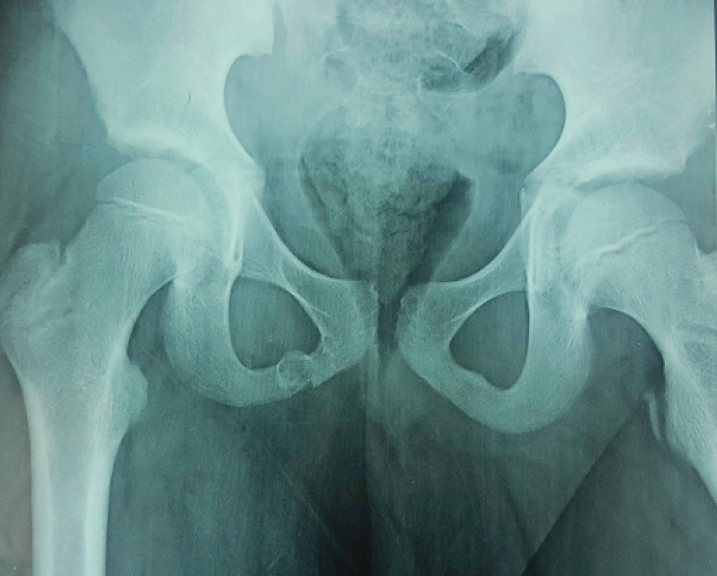
            Figure 1.

            X-ray showing lytic lesion in the right inferior ischio-pubic ramus with area of lucencies.
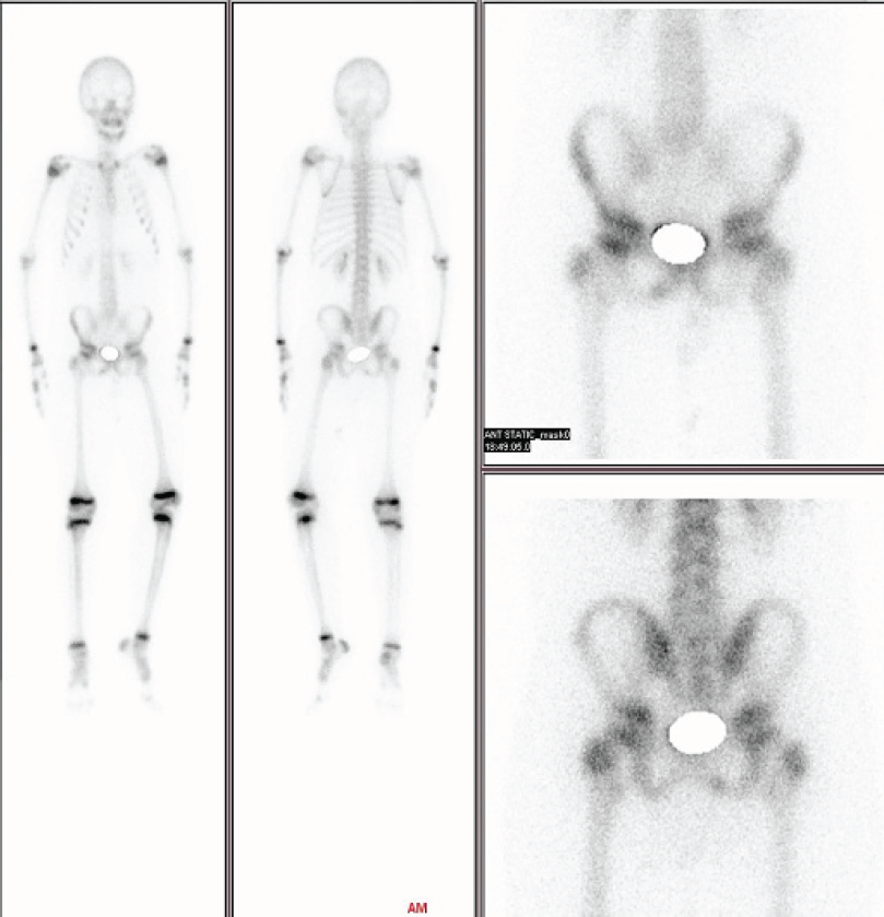
            Figure 2.

            99mTc-MDP bone scan showing focal area of increased uptake in the right inferior ischio-pubic ramus.
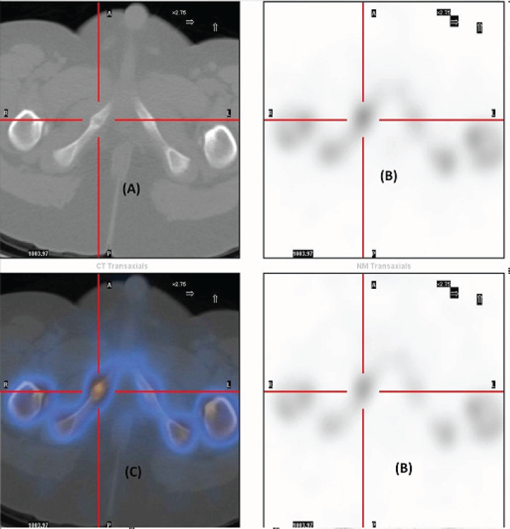
            Figure 3.

            99mTc-MDP bone scan with SPECT/CT images. (A) CT images showing areas of demineralization at edges of ischio-pubic synchondrosis and areas of sclerosis. (B) SPECT showing focal increased uptake in the inferior ischio-pubic ramus. (C) Fused SPECT/CT images confirms focal uptake to be in ischio-pubic synchondrosis.

            Discussion

            Ischio-pubic synchondrosis (IPS) is a transitory hyaline cartilage between inferior ischial and pubic rami [1]. This joint usually obliterates completely before puberty by bony union [2]. Its enlargement is not an uncommon radiological finding due to asymmetric closure of IPS, though it remains clinically asymptomatic. In some cases, children may present with pain in the groin, hip or gluteal region with restriction of hip movement and limping gait. This combination of clinical symptoms is usually referred as Synchondrosis Ischio-pubic syndrome (SIS) [3,4]. On plain X-rays, SIS is outlined as a bulge with areas of demineralization at IPS, referred by Odelberg and van Neck as “osteochondritis ischio-pubica” or “Van Neck-Odelberg disease” [5,6]. Multiple factors lead to enlargement of IPS, by inducing inflammatory response leading to delayed/deranged ossification. Most important of all, is uneven physical strain like kicking, dancing or jumping [4]. On conventional radiographs, its tumor like appearance can create panic. Therefore, it is very important to correctly and timely diagnose Ischio-pubic osteochondritis (IPO) as a benign disorder. Computed tomography (CT) scan usually show areas of sclerosis and lucencies. Diagnosis on magnetic resonance imaging (MRI) is usually on basis of abnormal marrow signals with fibrous bridging [4]. The differential diagnosis includes stress fracture, post traumatic osteolysis, osteomyelitis, and tumor [7,8]. Stress fractures of pubic rami in athletes can be challenging at times. Osteomyelitis is usually associated with a cascade of clinical symptom. Radiologically bony destruction with adjacent soft tissue collection is usually a hallmark of osteomyelitis. A 99mTc-MDP bone scan is widely used for evaluation of osteoblastic bone lesions [9]. Osteo-blastic bone lesions can be identified about 18 months earlier on bone scan than X-rays [10]. Bone scan is least sensitive for lytic lesions. Multiple benign focal pathologies of bone can lead to false positive findings of skeletal metastasis/tumors e.g. fibrous dysplasia, enchondroma, osteoid osteoma, brown tumors due to hyper-parathyroidism, and degenerative bone disease [11]. Planer 99mTc-MDP bone scan with additional SPECT/CT hybrid imaging can enhance the sensitivity and specificity of procedure to pick small lesions and effectively characterize the target lesion [12,13]. In our case SPECT/CT helped to localize the target lesion to be in IPS, with additional characterization of the lesion with CT images. Correlating clinical picture with 99mTc-MDP bone scan and SPECT/CT images, a diagnosis of Ischio-pubic Osteochondritis (Van Neck-Odelberg disease) was made. Latter after treatment by orthopedic surgeon with anti-inflammatory, steroids and analgesic drugs coupled with complete bed rest improved symptoms dramatically, confirming diagnosis of Ischio-pubic Osteochondritis.

            Conclusion

            Ischio-pubic Osteochondritis (Van Neck-Odelberg disease) is one of the differential diagnosis (DDs) in children presenting with pain in the groin, hip or gluteal region and limping gait. It is emphasized that ample awareness of this ailment is necessary for accurate and timely diagnosis in children. Early diagnosis and proper management can alleviate the suffering of child.

            Acknowledgements

            The authors would like to acknowledge the efforts and help of all the staff in the Department of Nuclear Medicine in preparation of this case report.

            List of Abbreviations

            CT

            Computed Tomography

            IPO

            Ischio-Pubic Osteochondritis

            IPS

            Ischio-Pubic Synchondrosis

            MDP

            Methylene Di Phosphonate

            MRI

            Magnetic Resonance Imaging

            SIS

            Synchondrosis Ischio-pubic Syndrome

            SPECT

            Single Photon Emission Computed Tomography

            Conflicts of Interest

            No conflict of interest.

            Funding

            None

            Consent from patient

            Informed written consent was obtained from the patient to publish this case report.

            Ethical approval

            Approved by local ethical committee of institute.

            Author’s Contribution

            MI is the main author of the manuscript. MN helped in the write up. MN and MSA helped in literature review. MBI critically analyzed the manuscript and helped in writing discussion.

            References:

            1. Macarini Luca, Lallo Tania, Milillo Paola, Muscarella Silvana, Vinci Roberta, Stoppino Luca P. Case report: Multimodality imaging of Van Neck-Odelberg disease. Indian J Radiol Imaging. 2011. Vol. 21(2):107–110

            2. Ceroni D, Mousny M, Anooshiravani-Dumont M, Buerge-Edwards A, Kaelin A.. MRI abnormalities of the ischiopubic synchondrosis in children: A case report. Acta Orthop Belg. 2004. Vol. 70:283–6

            3. Herneth AM, Philipp MO, Pretterklieber ML, Balassy C, Winkelbauer FW, Beaulieu CF.. Asymmetric closure of ischiopubic synchondrosis in pediatric patients: correlation with food dominance. AJR. 2004. Vol. 182:361–65

            4. Herneth AM, Trattnig S, Bader Tr, Ba-Ssalamah, Ponhold W, Wandl-Vergesslich K, Steinbach LS.. MR imaging of the ischiopubic synchondrosis. Magn Reson Imaging. 2000. Vol. 18:519–24

            5. Olderberg A.. Some cases of destruction in the ischium of doubtful etiology. Acta Chir Scand. 1924. Vol. 56:273

            6. Van Neck M.. Osteochondrite du pubis. Arch Franco-Belg Chir. 1924. Vol. 27:238–41

            7. Keats TE.. Plain film radiography: sources of diagnostic errors. Resnick D. Diagnosis of bone and joint disorders. 3rd. San Diego: Saunders. 1996. p. 41–67

            8. Oliveira Fillipe. Differential diagnosis in pain full IPS –a case report. Lowa Orthop J. 2010. Vol. 30:195–200

            9. Mettler FA, Milton JG. Skeletal system, Essentials of Nuclear Medicine Imaging. 6th ed. Philadelphia, PA: Elsevier/Saunders. 2012. 271–314

            10. Choi J Raghavan M.. Diagnostic imaging and image-guided therapy of skeletal metastases. Cancer Control. 2012. Vol. 19(2):102–12

            11. O’Sullivan Gerard J, Carty Fiona L, Cronin Carmel G. Imaging of bone metastasis: An Update. World J Radiol. 2015. Vol. 7(8):202–211

            12. Even-Sapir E, Flusser G, Lerman H, Lievshitz G, Metser U.. SPECT/multislice low-dose CT: a clinically relevant constituent in the imaging algorithm of nononcologic patients referred for bone scintigraphy. J Nucl Med. 2007. Vol. 48:319–24

            13. Saha S, Burke C, Desai A, Vijayanathan S, Gnanasegaran G.. SPECT-CT: applications in musculoskeletal radiology. Br J Radiol. 2013. Vol. 86:20120519

            Summary of the case

            Patient, age/gender 111 year/Male
            Final Diagnosis 2Ischio-pubic Osteochondritis (Van Neck-Odelberg disease)
            Symptoms 3Pain in the right hip
            Medications (Generic names only) 4NSAIDs, steroids
            Clinical Procedure 5X-ray Pelvis, 99m-Tc-MDP bone scintigraphy, SPECT/CT hybrid imaging.
            Specialty 6Nuclear Medicine
            Objective 7To diagnose cause of pain in the right hip.
            Background 8Ischio-pubic synchondrosis, Ischio-pubic osteochondritis, 99m-Tc-MDP bone scintigraphy, SPECT/CT.
            Case Report 9Ischio-pubic Osteochondritis (Van Neck-Odelberg disease) – A case report
            Conclusion 10Ischio-pubic Osteochondritis – Important to diagnosis it early for effective treatment.
            MeSH Keywords 11 99mTc-MDP bone scintigraphy, SPECT/CT, Ischio-pubic Osteochondritis, Van Neck-Odelberg disease, Case report

            Author and article information

            Journal
            European Journal of Medical Case Reports
            EJMCR
            Discover STM Publishing Ltd.
            2520-4998
            30 September 2017
            : 1
            : 3
            : 144-147
            Affiliations
            [1]Department of Nuclear Medicine, PINUM Cancer Hospital, P.O. Box: 2019, Jail road, Faisalabad, Pakistan
            Author notes
            [* ] Correspondence to: Muhammad Iqbal Nuclear Medicine Physician, PINUM Cancer Hospital, P.O. Box 2019, Faisalabad,Pakistan Email: Iqbalpinum@ 123456gmail.com
            Article
            ejmecr-1-144
            10.24911/ejmcr/1/34
            6a3b4571-17ae-4140-9b4e-2ba116720695
            © Muhammad Iqbal, Muhammad Naeem, Muhammad Babar Imran, Muhammad Shahzad Afzal, Muhammad Saeed Akhtar

            This is an open access article distributed in accordance with the Creative Commons Attribution (CC BY 4.0) license: https://creativecommons.org/licenses/by/4.0/) which permits any use, Share — copy and redistribute the material in any medium or format, Adapt — remix, transform, and build upon the material for any purpose, as long as the authors and the original source are properly cited.

            History
            : 28 March 2017
            : 25 August 2017
            Categories
            CASE REPORT

            99mTc-MDP bone scintigraphy,Ischio-pubic Osteochondritis,case report,Van Neck-Odelberg disease,SPECT/CT

            Comments

            Comment on this article