Background
Fibroadenomas are benign lesions in the breast and occurs around 2% in adolescent females. This is found in 68% of all the breast masses and 10-15% may be multiple in nature [1]. Though these masses remain asymptomatic but these can cause pain as the size increases.25 to 40 years of age is the peak incidence period of their occurrence. The entity may be associated with Cowden syndrome where multiple hamartomas are present.
Case presentation
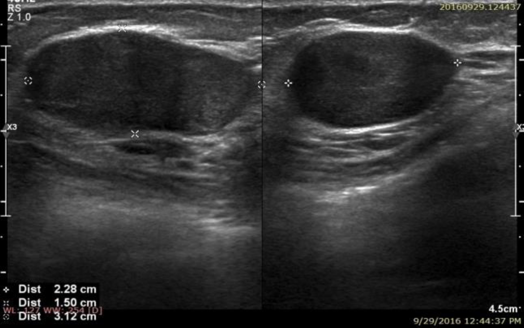
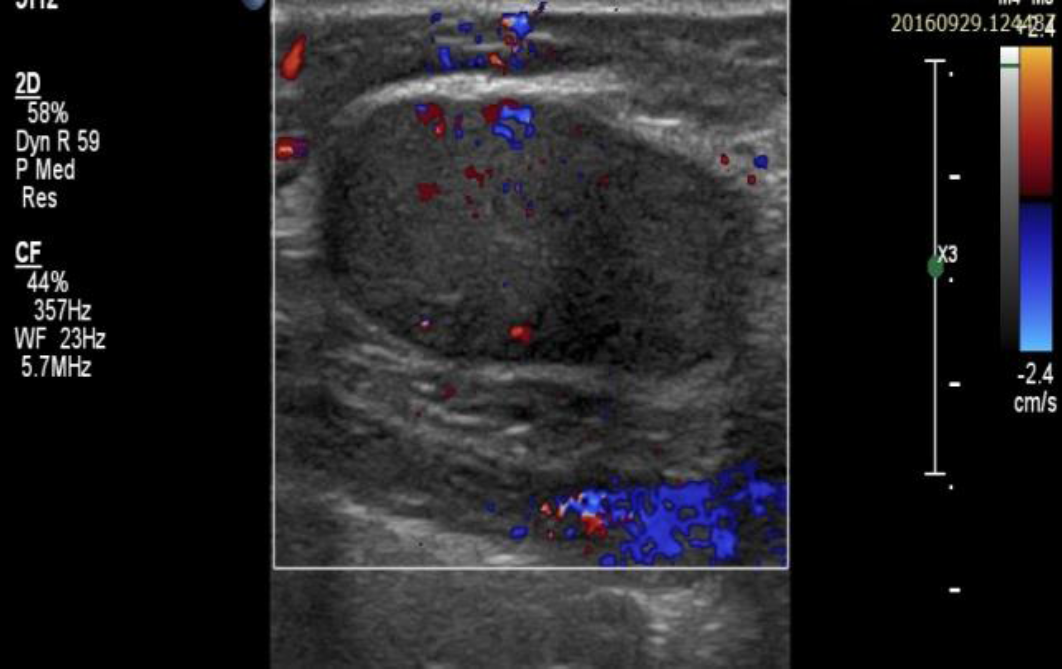
A 21-years old female presented with palpable mass in the left breast for over one year duration. This became slightly painful off and on, and more during monthly cycles. There was no increase in size of the lump did not increase in this period. Patient was having anxiety for having this lump in the breast. There was no family history of this disease. There was no history of any previous interventional procedure. The examination of the breast revealed palpable mass on the outer quadrant of left breast which was slightly tender and measured approximate 3 cm x 2.5 cm. Rest of the systemic examination was within normal limits. Biochemical parameters were also within normal limits. Ultrasonogrpphy of left breast revealed a hypoechoic mass in left breast. These masses did not have any cystic component and there was no evidence of any calcification (Figure 1). Color flow imaging did not reveal much vascularity (Figure 2). Color doppler spectrum was normal. Ultrasonography of right breast was unremarkable.
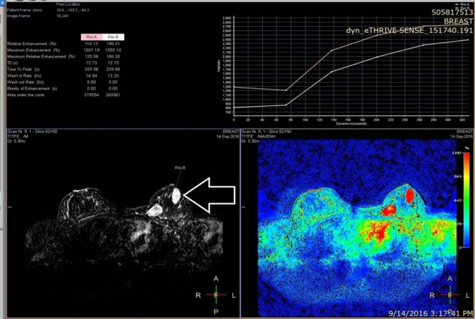
Magnetic resonance study has shown two lesions in the left as well as one lesion in the right breast. These were hypointense on T1W and hyperintense on STIR (short tau inversion recovery) sequences (Figure 3 and Figure 4). Dynamic contrast study was carried out which revealed classical delayed wash out pattern denoting Type 1 enhancement curve (Figure 5 and Figure 6).
Histopathological biopsy confirmed the diagnosis of benign fibroadenomas. The specimen had fibroblastic stroma having glandular and cystic spaces lined with luminal epithelial and outer myoepithelial cells. The patient has been put on regular three months monitoring and follow up as the patient is young without any complications. The decision for surgical management will be taken if the tumor increases in size on the grounds of psycho-social-cosmetic reasons.

Ultrasonogrphic image of the left breast. There is well defined hypoechoic lesion in the outer upper quadrant which measures 3.12 x 2.28 x 1.5 cm. There is no calcification or necrosis seen.

Color flow imaging of the left breast lesion. There was very less vascularity observed in the lesion. The margins were also well defined without any abnormal vessels.

MR of both breasts T1W sequences. (a) plain image shows isointense (white solid arrow) lesion in the left breast indistinguishable from the surrounding tissue. (b) T1W image shows moderate enhancement (white horizontal hallo arrows) after gadolinium injection. The outer lesion shows a few septations.

STIR sequences. (a) Both the breasts show well defined hyperintense lesions (white hollow vertical arrows). (b) Left breast shows another anterior lesion with the same morphological features.

Dynamic contrast study shows the pattern of the contrast enhancement graph in the superficial lesion (horizontal arrow) of the left breast. This shows slow initial enhancement but delayed wash out (Type 1 curve). The curve is drawn as the ratio of maximum intensity change divided by the time interval.
Discussion
Fibroadenoma of the breast is an admixture of epithelial and stromal tissues. These tumors are often called as breast mice because of their mobility in the breast tissue [2]. This should not be confused with fibroadenosis. The entity has to be differentiated from other lumps as the carcinoma can also present in the similar fashion. These are divided into two categories as per their stroma and epithelial cell components as pericanalicular and intracanalicular. These have got intact and compressed glandular spaces respectively. There is proliferation of the stroma in the latter category. Fibroadenoma masses are easy to be removed as compare to the malignant pathologies. These present as solitary firm, mobile, painless, rubbery and slowly growing mass. Sometimes these can be of large size representing as giant fibroadenoma [3]. These occur during the child bearing period and may regress after menopause. These can be of multiple in nature and can occur bilaterally [4]. This is less prevalent in males because of the hormonal reasons. These arise from lobular stroma of the terminal duct lobular unit. These can be confused with phyllodes tumor as the site of occurrence is same. The exact etiopathology and causative factors of fibroadenomas are not known. Their occurrence is partially related to the sensitivity to the female estrogen hormone [5]. There is some evidence of genetically related to the family history for the occurrence. These are more common during fertility period of females and may regress after menopause [6]. The choice of investigating modality is as per age. Dynamic contrast MR study shows classical delayed wash out graph. This had been depicted in our case also in both the superficial as well as deep lesions. The younger age group can undergo ultrasound and Magnetic resonance imaging examinations but the patients older than 35 years Mammography is advised in addition to other choices [7]. Contrast MR studies are able to distinguish the benign and malignant masses on the basis of shape, outline, lobulation and internal septation. Benign and malignant pathologies were differentiated by the ratio of maximum intensity change divided by the time interval during which the first occurred. There is so much overlap which requires histopathological confirmation [8]. Regular follow-up and monitoring is best advised [9, 10]. In some cases, surgical excision is required and the resection is not that difficult as the margins are very clear. If phyllodes tumor is there, then the margins have to include some part of the breast tissue [11]. Cryotherapy and radiofrequency ablation are the alternative management methods if the patients are young. Regular follow-up at different intervals is done for the evaluation [12, 13].
Conclusion
Solitary or multiple adolescent fibroadenomas are not uncommon. The diagnosis is very important and great accuracy is achieved with ultrasound coupled with mammography. Mammography should be avoided in adolescent patients. Contrast enhanced dynamic MRI scanning provides strong tool to differentiate fibroadenoma from the malignant pathologies as was in our present case. In majority of cases, monitoring and follow up is done rather than contemplating surgical approach. If the lesion is very big and painful then surgical excision is the answer.


