129
views
0
recommends
+1 Recommend
1 collections
    0
    shares
      scite_
      0
      0
      0
      0
      Smart Citations
      0
      0
      0
      0
      Citing PublicationsSupportingMentioningContrasting
      View Citations

      See how this article has been cited at scite.ai

      scite shows how a scientific paper has been cited by providing the context of the citation, a classification describing whether it supports, mentions, or contrasts the cited claim, and a label indicating in which section the citation was made.

       
      • Record: found
      • Abstract: found
      • Article: found
      Is Open Access

      Monorchism cryptorchidism: what should be the imaging evaluation protocol? - a case report

      Published
      case-report
      Bookmark

            Abstract

            Background:

            Monorchism is having only one testis and cryptorchidism is undescended testis. This is usually congenital anomaly but can happen as a result of trauma or surgical procedure undertaken. There are great psychosocial factors related to this entity. The complete evaluation is of paramount importance. Radiological protocol for detailed search for the hidden testis is of utmost importance for future management procedure.

            Case presentation:

            We present 2-years old male child who was evaluated for having empty scrotal sac. He underwent Ultrasound (US), and magnetic resonance imaging (MRI) for the evaluation. Diffusion weighting images (DWI) played a great role in the diagnosis. The child was having only left sided testis and that too was undescended.

            Conclusion:

            There is great value of cross sectional imaging in diagnosing the hidden testis. The sonography remains the first investigation of choice. MRI should be undertaken to locate the testis but may fail also for the localization. Diffusion weighted imaging (DWI) plays an important role with great reliability in the diagnosis among pediatric cases being radiation free modality.

            Main article text

            Background

            Cryptorchidism is the absence of one or both testicles from the scrotal sac. This is a Greek word which means hidden testicle. The incidence is higher in premature to 30% as compared to a full term being 3%. The incidence is 1-6% of male children. This is called as monorchism if only one testis is present. Monorchism cryptorchidism is single undescended testis. Ectopic testis differs from this as in that case there is deviation of the testicle from the normal descent path and can lie somewhere else. The entity should also not be confused with retractile testis which is a normal phenomenon.

            Case presentation

            A 2 year old male child (Figure 1) was brought to children outpatient department by the parents with missing testes in the scrotal sac. The child was of full term delivery without any complications.

            On examination, the child physical parameters were as per his age. Systemic examination was unremarkable. On local examination, it was found that the scrotal sac was empty as testes were not palpable. All the blood investigations were within normal limits. Plain skiagram for abdomen did not reveal anything. The visible bone structures were normal. The child underwent ultrasound (US) study which revealed empty scrotal sac without any anything within it. There was another well-defined testis-like hypoechoic structure seen in the left inguinal region. No other finding was noticed in the vicinity or on the contra lateral side (Figure 2).

            Color Doppler did not show any flow in the testis and the surrounding region was unremarkable (Figure 3). The child was planned for magnetic resonance study of the abdomen. MRI study revealed a mixed intensity mass in T2W on the left inguinal region but there was no such structure seen on right side. T1W images were equivocal. Diffusion weighted imaging (DWI) had revealed a well-defined hyper intense structure in the left inguinal region. This is the same structure which was seen in T1W sequences (Figures 4 a, b,c, 5 a, b and 6).

            The child was diagnosed with absence of right testis with single left undescended testis. The hormonal estimation had been sent and the child is planned for orchidopexy.
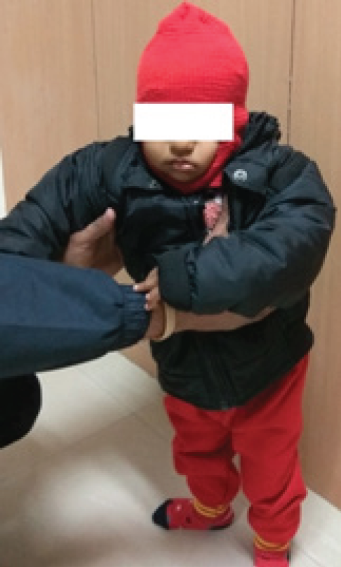
            Figure 1:

            Photo of the two year old male child with normal height, weight and other physical parameters.
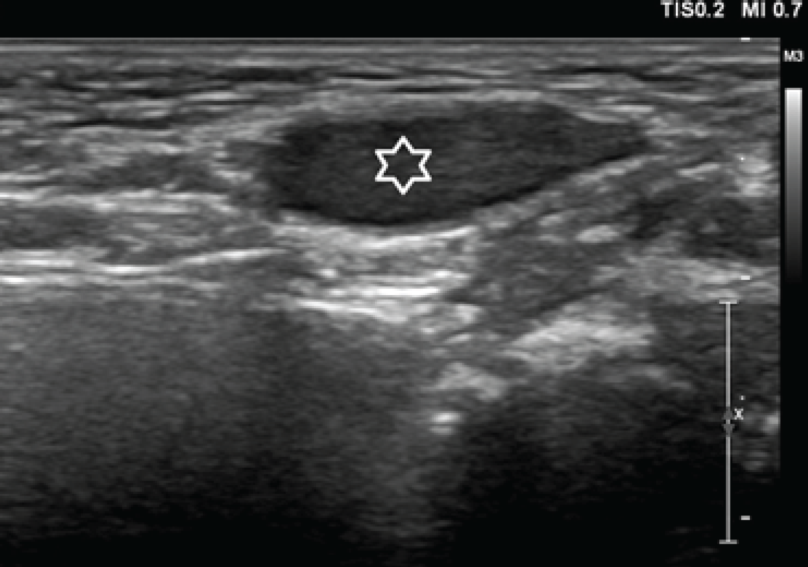
            Figure 2:

            Ultrasound of the left inguinal region with 7.5 MHz linear probe. There is a well-defined oval hypoechoic region which is suspicious of left testis (white star).
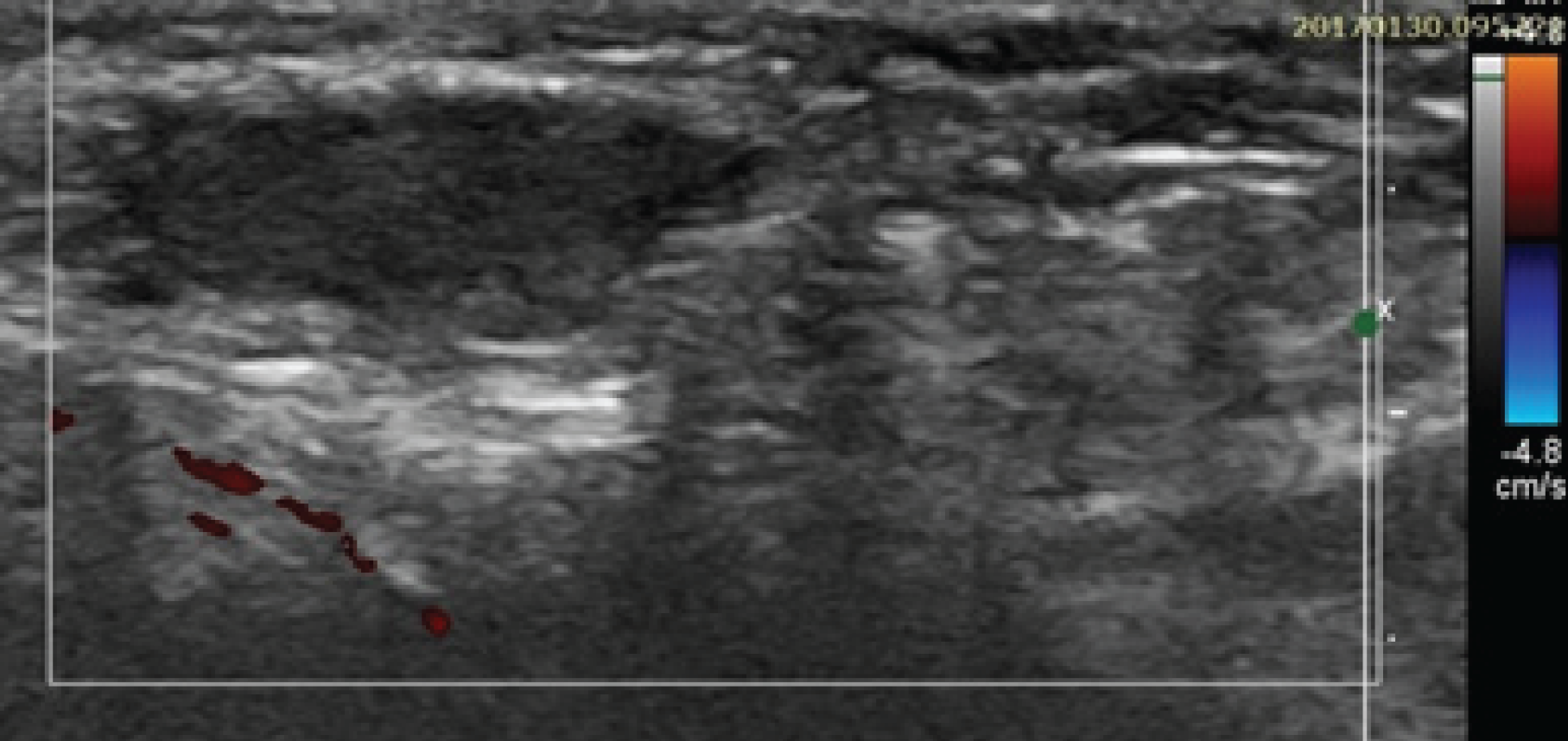
            Figure 3:

            Color Doppler of the left testis in the inguinal canal region did not reveal any vascularity and the margins were very clear.

            Figure 4:

            Axial sections of magnetic resonance imaging study. (a) T2W image at the level of left testis in inguinal canal (inverted white arrow). The testis is slightly hyperintense comparing to muscles. (b) T2W at slightly lower level shows only white intensity of the inguinal canal (white hollow arrow). (c) Balanced Turbo Field Echo (BTFE) image shows testis of hyperintensity in the canal (white arrow).

            Figure 5:

            MRI axial sections. (a) BTFE image shows hypointense structure in the left inguinal canal (thin white inverted arrow), (b) Diffusion weighted image (DWI) shows same structure as markedly hyperintense (inverted thick white arrow).

            Figure 6:

            Coronal T2W section of abdomen shows bilaterally normally placed kidneys without any structural abnormality.

            Discussion

            Monorchism cryptorchidism is the presence of single undescended testis. Scrotal sac is usually empty. The embryonic development of testis starts at the 10th week of gestation in the abdominal cavity and comes to lie in inguinal canal and further to scrotal sac between 28th to 40th weeks of gestation. The gubernaculum (Latin word which means rudder) ligament under the influence of testosterone hormone contracts and brings the testes in the scrotal sac. In general, 60% cases shows normal descend of both the testes in scrotal sac by the end of one year. There can be various permutation and combinations during this descent and child can present accordingly with various congenital entities.

            In 72% cases, the cryptorchid testis lies in inguinal canal as in our case, 20% lies in prescrotal and only 8% are seen in intra-abdominal location. The chance of testicular descent after one year is unlikely. Our case was having only one testis and that too undescended, lying in inguinal canal. There are more chances of 20-48 times for malignant transformation. There is an incidence of 5-10% undescended testis in testicular cancers [3]. This is also one of the causes of infertility and sometimes leads to hernia. Cross-sectional Imaging plays a pivotal role in locating the undescended testis. Ultrasonography (US), computerized tomography (CT), magnetic resonance imaging (MRI) and arteriography along with laparoscopy are the methods to find out cryptorchid testis. Ultrasonography remains the first choice to locate the testis below internal inguinal ring. This is noninvasive and without any radiation.

            The testis is seen as oval region of low or midlevel echogenicity. This has got 91% accuracy [4]. Color Doppler shows low resistance flow spectrum in the testis [5,6]. CT is avoided because of radiation issue; otherwise this scores over US in differentiating testis from surrounding lymph node. This has got 96% accuracy. MRI is of great importance because of better tissue characterization, multiplanar capabilities, noninvasive and without any radiation. Testis is seen as hypointense in T1W and hyperintense on T2W images. DWI shows markedly Hyperintensity which is characteristic of the testis [7,8].

            Orchidopexy is the choice of management [9,10]. The main aim of the surgery is to bring back the testicle to the normal position, to preserve hormonal function, fertility and to avoid future malignancy [11]. This is performed from 5 to 15 months of age. Hormonal therapy is also helpful to some extent but the results are not encouraging. Surgery should be performed in the first year otherwise, this will lead to loss of testicular functions and the condition is labeled as vanishing testis.

            Conclusion

            Undescended testes pose a great diagnostic challenge for their localization. The diagnosis is important because of the fear of malignant potential. The radiation should be avoided in children. Though ultrasound is the first screening modality of the diagnosis but DWI in MRI has got more diagnostic value.

            Acknowledgements

            We are thankful to Mr. Nitish Virmani, Mr. Rajdeep Thidwar, Miss Nikita Khanna and Mr. Raushan Singh of Faculty of Allied Health Sciences of SGT University for carrying out all the investigations and preparing the manuscript.

            List of Abbreviations

            CT

            Computerized tomography

            DWI

            Diffusion weighted imaging

            MRI

            Magnetic Resonance Imaging

            Conflict of Interests

            None

            Funding

            None

            Consent for publication

            Informed consent was obtained from the parents to publish this case in a medical journal.

            Ethical approval

            Ethical approval is not required at our institution for publishing a case report in a medical journal.

            Authors’ contribution

            BB wrote the first draft of the manuscript. SS provided clinical details of the case, NB provided the data and helped in writing the manuscript. SD provided data. MRA provided US and MR images. SS carried out MRI investigation. All authors approved the final version of the paper.

            References

            1. Snodgrass W, Bush N, Holzer M, Zhang S.. Current referral patterns and means to improve accuracy in diagnosis of undescended testis. Pediatrics. 2011. Vol. 127(2):e382–388

            2. Niedzielski JK, Oszukowska E, Słowikowska-Hilczer J.. Undescended testis –current trends and guidelines: a review of the literature. Arch Med Sci [Internet]. 2016. 2017 Feb 28Vol. 12(3):667–77. http://www.ncbi.nlm.nih.gov/pmc/articles/PMC4889701

            3. Swerdlow AJ, Higgins CD, Pike MC.. Risk of testicular cancer in cohort of boys with cryptorchidism. BMJ. 1997. Vol. 314(7093):1507–11

            4. Dogra VS, Gottlieb RH, Oka M, Rubens DJ.. Sonography of the scrotum. Radiology. 2003. Vol. 227(1):18–36

            5. Bree RL, Hoang DT.. Scrotal ultrasound. Radiol Clin North Am. 1996. Vol. 34(6):1183–205

            6. Krone KD, Carroll BA.. Scrotal ultrasound. Radiol Clin North Am. 1985. Vol. 23(1):121–39

            7. Krishnaswami S, Fonnesbeck C, Penson D, McPheeters ML.. Magnetic resonance imaging for locating nonpalpable undescended testicles: a meta-analysis. Pediatrics. 2013. Vol. 131(6):e1908–1916

            8. Emad-Eldin S, Abo-Elnagaa N, Hanna SA, Abdel-Satar AH.. The diagnostic utility of combined diffusion-weighted imaging and conventional magnetic resonance imaging for detection and localization of non palpable undescended testes. J Med Imaging Radiat Oncol. 2016. Vol. 60(3):344–51

            9. Kirsch AJ, Escala J, Duckett JW, Smith GH, Zderic SA, Canning DA, et al.. Surgical management of the nonpalpable testis: the Children’s Hospital of Philadelphia experience. J Urol. 1998. Vol. 159(4):1340–3

            10. Tsujihata M, Miyake O, Yoshimura K, Kakimoto K, Matsumiya K, Takahara S, et al.. Laparoscopic diagnosis and treatment of nonpalpable testis. Int J Urol. 2001. Vol. 8(12):692–6

            11. Hadziselimovic F, Herzog B.. The importance of both an early orchidopexy and germ cell maturation for fertility. Lancet. 2001. Vol. 358(9288):1156–7

            Summary of the case

            Patient (gender, age) 1Male, 2 year old
            Final Diagnosis 2Monorchism cryptorchidism
            Symptoms 3Empty scrotal sac
            Medications (Generic) 4NIL
            Clinical Procedure 5Ultrasound and MRI
            Specialty 6Pediatrics
            Objective 7To find out the location of undescended testis
            Background 8Child with empty scrotal sac
            Case Report 9Cross-sectional imaging for Cryptorchidsm in Pediatric age group
            Conclusions 10DWI of MRI is conclusive and diagnostic in hidden testes.
            MeSH Keywords 11Monarchism, cryptorchidism, psychosocial, US, MRI, DWI, case report

            Author and article information

            Journal
            European Journal of Medical Case Reports
            EJMCR
            Discover STM Publishing Ltd.
            2520-4998
            30 May 2017
            : 1
            : 2
            : 92-96
            Affiliations
            [1 ]Department of Radio-diagnosis, SGT Medical College Budhera (Gurgaon), India.
            [2 ]Department of Pediatrics, SGT Medical College Budhera (Gurgaon), India.
            [3 ]Faculty of Allied Health Sciences, SGT University Gurgaon (Haryana), India.
            Author notes
            [* ] Correspondence to: Dr. Bharat Bhushan Sharma, Professor & HOD, Department of Radio-diagnosis, SGT Medical College, Budhera, (Gurgaon) 122505, India. Email: bbhushan986@ 123456gmail.com
            Article
            ejmcr-1-92
            10.24911/ejmcr/1/18
            ed04b19f-4224-4626-8084-2dbb10951ba6
            © Bharat Bhushan Sharma, Shashi Sharma, Naveen Bhardwaj, Sakshi Dewan, Mir Rizwan Aziz, Shilpa Singh

            This is an open access article distributed in accordance with the Creative Commons Attribution (CC BY 4.0) license: https://creativecommons.org/licenses/by/4.0/) which permits any use, Share — copy and redistribute the material in any medium or format, Adapt — remix, transform, and build upon the material for any purpose, as long as the authors and the original source are properly cited.

            History
            : 08 February 2017
            : 01 March 2017
            Categories
            CASE REPORT

            US,psychosocial,cryptorchidism,monarchism,case report,DWI,MRI

            Comments

            Comment on this article