Background
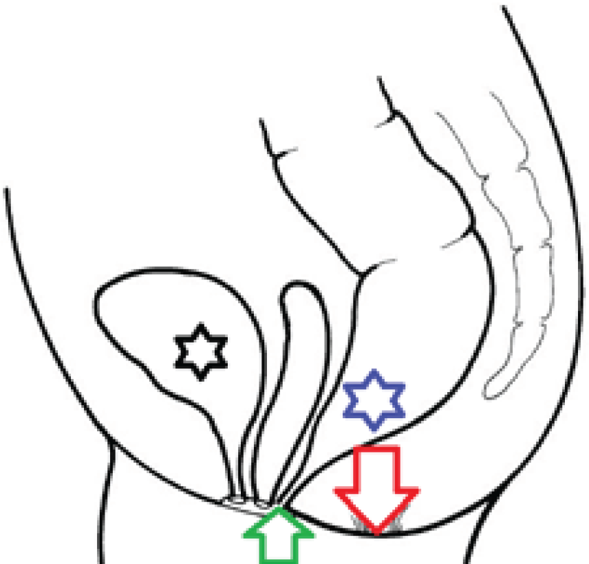
Rectovestibular fistula (RVF) is the communication between rectum and vulvar vestibular part which lies posterior to the hymen. The ectopic anal opening lies in the frenulum of the labia minora. It is important to know the exact anatomical locations of all the orifices in the perineum. In our case, there was imperforate anus and there was common opening for the vagina and rectum (Figure 1).
Case presentation
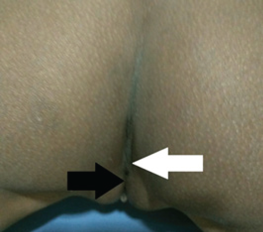
A 7 year old female child was brought to the pediatric outpatient department with the history of passing feces and gas through vaginal opening since birth (Figure 2).
There was also history of recurrent urinary tract infection and sometimes passing pus like material from the opening. The child was born in a remote rural region without any accessible obstetric or pediatric assistance. There was no proper anal orifice at birth. On examination, the child was found to be of average physical parameters without any abnormality in systemic examination.
Local examination of the external genitalia and perineum revealed two openings as that of urethra and vagina along with another small fistulous opening near the vaginal orifice. There was pus like material oozing out of the vaginal orifice along with surrounding inflammation. Anal orifice was absent (Figure 3).
Vital parameters of the child were preserved. The consent of the parents was taken for the investigations of their child. Ultrasound examination was unremarkable and there was no other internal congenital anomaly. MRI of pelvis was done which showed the anatomical detailed relationship (Figure 4).
There was common opening of rectum and vagina at the perineum. Conventional study was carried out after injecting the contrast in the vaginal orifice which revealed the free flow of the contrast in the rectum (Figure 5). The diagnosis of recto-vestibular fistula was confirmed with imperforate anus. The child has been planned for pre surgical colostomy which is to be followed by posterior sagittal anorectoplasty (PSARP).

Sagittal diagrammatic representation of the rectovestibular fistula in the pelvis and perineal region. Anal orifice is absent at the normal place (red downward arrow) and rectum is opening along with vaginal orifice (green upwards arrow). Urinary bladder (black star) lies anteriorly and rectum on the posterior most location (blue star).

Photograph of perineum and external genitalia. There is normal urethral orifice on anterior aspect (black arrow) with posteriorly common passage of the vagina and rectum (white arrow).
Discussion
Anorectal malformation forms a complex group of anomalies where the baby is born with imperforate anus [1]. Rectum may open at different location leading to different types of fistulous communications. Congenital rectovestibular fistula (CRF) is one of these anomalies. Rectovestibular fistula is found exclusively in females and is of congenital in nature [2]. This is often confused with rectovaginal fistula which is found in less than 1% cases [3]. There is no definitive causative factor noted in this anomaly but the drugs used during pregnancy are thought to be one of the reasons for this anomaly. This may be associated with the sacral spinal or renal anomalies. Anorectal malformations are associated with VACTERL (vertebral, anus, cardiac, trachea, esophagus, renal and limb) anomalies [4].
Radiological evaluation plays a great role in diagnosing these anomalies by different modalities. Plain Xray can tell the details of the bony abnormalities. US (Ultrasound) and MRI further evaluate the rest of associated anomalies. Conventional studies with contrast will exactly highlight the fistulous communication which will decide the course of management. Our case was also diagnosed by injecting contrast in the fistulous opening delineated the communication to the rectum. Three stage surgery would be performed. The surgical management is warranted after the colostomy. Posterior sagittal anorectoplasty (PSARP) is performed as a choice [5–7]. There is always fear of stool incontinence because of the anal sphincter problems.


