63
views
0
recommends
+1 Recommend
1 collections
    0
    shares
      scite_
      0
      0
      0
      0
      Smart Citations
      0
      0
      0
      0
      Citing PublicationsSupportingMentioningContrasting
      View Citations

      See how this article has been cited at scite.ai

      scite shows how a scientific paper has been cited by providing the context of the citation, a classification describing whether it supports, mentions, or contrasts the cited claim, and a label indicating in which section the citation was made.

       
      • Record: found
      • Abstract: found
      • Article: found
      Is Open Access

      Type II right sided aortic arch with aberrant left subclavian artery (ALSA) – what clinician wants to know? - a case report

      Published
      case-report
      Bookmark

            Abstract

            Background:

            Right sided aortic arch is slightly rare variant of aortic arch which requires more description and elaboration for the management in clinician point view. This may remain asymptomatic or present with various vague symptomatology. It is mandatory for the clinician to be aware of the underlying anatomical details which may sometimes be responsible for the presenting complaints. Furthermore, this anomaly may also be associated with other abnormalities.

            Case presentation:

            We present 60-years old male who complained of headache and vague chest pain of two weeks duration and underwent Contrast Enhanced Computerized Tomography (CECT) chest for further evaluation. There was an incidental finding of right sided aortic arch with Aberrant Left Subclavian Artery (ALSA). This anomaly was not responsible for any of the symptoms as per the CECT findings.

            Conclusion:

            It is important to know the detailed relationship of right sided aortic arch with ALSA to the other neighboring anatomical structures. MDCT and MRI are always helpful tools to appreciate and apprise the clinician about the details of the anomaly in view of the management if required. Sometimes the symptoms may be present because of the compression by the vascular rings.

            Main article text

            Background

            Right sided aortic arch with aberrant left subclavian artery (ALSA) is seen in 0.05% of the general population. The incidence of isolated right aortic arch is approximate 0.1% [1]. Originally, only aberrant right subclavian having slight dilatation at the origin was called as Kommerell diverticulum. This was named after Burckhardt Kommerell in 1936. Now the dilatation of aberrant subclavian at the origin is also called by the same name [2].

            Case presentation

            60-years old male reported to otolaryngology department with complaints of mild headache with vague chest pain and neck pain of two weeks duration. On examination, the patient was of averagely built without any previous history of illness or any systemic disease. Systemic examination was unremarkable. X-ray paranasal sinus was unremarkable. Plain X-ray chest had shown right sided aortic arch without any other abnormality (Figure 1).

            All the basic blood investigations were within normal parameters. The patient underwent contrast-enhanced computerized tomography (CECT) with 16- slices Siemens Scope whole body CT scanner. The findings revealed right sided aortic arch with aberrant left subclavian artery (ALSA). There was no abnormality in both the lung fields and mediastinal anatomy was within normal limits. Aortic arch was seen encircling the trachea (Figure 2).

            The course of the thoracic aorta comes to the midline or to the left side as it approaches the diaphragmatic aperture (Figure 3). Common carotids on both sides could be seen originating from the arch independently
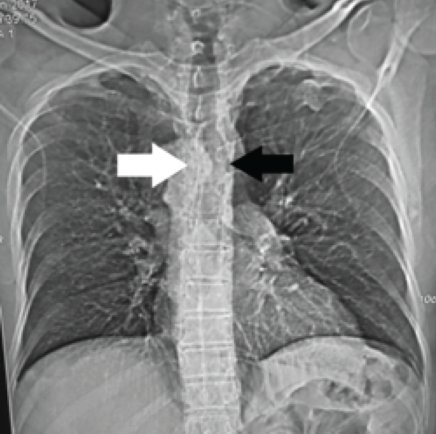
            Figure 1:

            Plain X-ray chest PA view. The aortic knuckle is seen on right side (white arrow) and there is tracheal indentation over trachea at the same level (black arrow). Rest of the mediastinal structures are at normal sites.
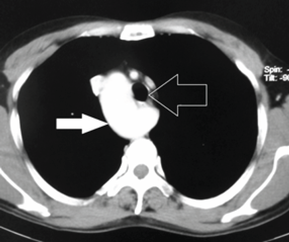
            Figure 2:

            CECT Thorax axial section at aortic arch level. Aortic arch is seen on right side (white arrow) with trachea on left side (hollow white arrow).

            Figure 3:

            CECT thorax at the diaphragm aperture. Aorta lies

            almost in midline position (hollow white arrow). (Figure 4) Left subclavian had taken origin from the right side and coursing towards the left side lying posterior to the esophagus (Figure 5).

            The patient was placed on symptomatic treatment as no further management was warranted in this case.

            Figure 4:

            CECT Chest 3D sagittal reformat image. Both common carotids are seen originating from the arch of the aorta (white arrow). Left common carotid is the first branch in order of the origin.

            Figure 5:

            Aberrant left subclavian artery (ALSA). (a) axial section shows the course of ALSA at upper part of the chest. (b) coronal reformat image shows aneurysmal origin (white star) with upward deviation to the left upper extremity.

            Discussion

            Isolated right sided aortic arch is comparatively a common vascular anomaly and in many times this can be visualized as an incidental finding in CECT chest. This develops because of the regression of the portion of the dorsal segment between the left subclavian and common carotid in the hypothetical double arch. There is also persistence of the fourth embryonic aortic arch on right side [3]. Sometimes this becomes symptomatic because of the vascular rings and due to other additional abnormal findings [4]. Following two types of entities are of clinical importance:

            Type I: Mirror image

            Type II: when present with aberrant left subclavian artery (ALSA)Our present case falls in Type II category. Type I is associated with other congenital anomalies. Type II is very rarely associated with congenital heart disease (CHD) as compared to Type I. The most common association with Type I is Tetrology of Fallot. ALSA can be retro-tracheal or retro-esophageal and may become symptomatic sometimes [5]. In our case, it was retro-esophageal and was asymptomatic. The order of origin of the vessels is left common carotid, right common carotid, right subclavian and then ALSA. This entity is slightly less common than the aberrant right side subclavian artery from the left side aortic arch. MDCT is the modality of choice for the delineation of these anomalies. Magnetic resonance imaging (MRI) and barium swallow play the additional role in the diagnosis of the vascular rings [6]. Dysphagia lusoria and dyspnea are the common complaints felt because of vascular rings. Magnetic Resonance Angiography (MRA) and Computerized Tomography Angiography (CTA) confirm the vascular mapping of the anomalies in detail.

            Conclusion

            Right sided aortic arch is diagnosed as an incidental finding in CECT chest as in our present case. The relationships of different structures are very important for the management point of view. CECT chest and angiography play an important role in these types of the cases.

            Acknowledgements

            We are thankful to Mrs. Shilpa Singh, Mr. Nitish Virmani, Mr. Rajdeep Thidwar, Miss Nitika and Mr. Raushan Singh of Faculty of Allied Health Sciences of SGT University Gurgaon (Haryana) for their contribution in carrying out the study and procuring the images.

            List of Abbreviations

            ALSA

            Aberrant left subclavian artery

            CECT

            Contrast-enhanced computerized tomography

            CHD

            Congenital heart disease

            CTA

            Computerized Tomography Angiography

            MRA

            Magnetic Resonance Angiography

            MRI

            Magnetic resonance imaging

            Conflict of Interests

            None

            Funding

            None

            Consent for publication

            Informed consent was obtained from the patient to publish this case in a medical journal

            Ethical approval

            Ethical approval was obtained from the ethical committee of the institute to publish this case report in a medical journal.

            Authors’ contribution

            BBS wrote the case report and supervised the project. SS carried out the clinical evaluation. NB helped in procuring the data. SD carried out cross-sectional imaging. MRA helped in collecting all the images for the study.

            References

            1. Türkvatan A, Büyükbayraktar FG, Olçer T, Cumhur T.. Multidetector computed tomographic angiography of aberrant subclavian arteries. Vasc Med. 2009. Vol. 14:5–11. [10.1177/1358863X08097903]

            2. Fisher RG, Whigham CJ, Trinh C.. Diverticula of Kommerell and aberrant subclavian arteries complicated by aneurysms. Cardiovasc Intervent Radiol. 2005. Vol. 28:553–60. [10.1007/s00270-003-0229-0]

            3. Lee EY, Siegel MJ, Hildebolt CF, Gutierrez FR, Bhalla S, Fallah JH.. MDCT evaluation of thoracic aortic anomalies in pediatric patients and young adults: comparison of axial, multiplanar, and 3D images. AJR Am J Roentgenol. 2004. Vol. 182:777–84. [10.2214/ajr.182.3.1820777]

            4. Shuford WH, Sybers RG, Gordon IJ, Baron MG, Carson GC.. Circumflex retroesophageal right aortic arch simulating mediastinal tumor or dissecting aneurysm. AJR Am J Roentgenol. 1986. Vol. 146:491–6. [10.2214/ajr.146.3.491]

            5. Dillman JR, Yarram SG, D’Amico AR, Hernandez RJ.. Interrupted aortic arch: spectrum of MRI findings. AJR Am J Roentgenol. 2008. Vol. 190:1467–74. [10.2214/AJR.07.3408]

            6. Hernanz-Schulman M.. Vascular rings: a practical approach to imaging diagnosis. Pediatr Radiol. 2005. Vol. 35:961–79. [10.1007/s00247-005-1529-0]

            Summary of the case

            Patient (gender, age) 1Male, 60 year old
            Final Diagnosis 2Right sided aortic arch with ALSA
            Symptoms 3Vague headache, chest and neck pain
            Medications (Generic) 4Symptomatic
            Clinical Procedure 5CECT Chest
            Specialty 6Radiodiagnosis and ENT
            Objective 7To find out the cause of SYMPTOMS
            Background 8Vague headache, chest and neck pain
            Case Report 9Cross-sectional imaging for the chest pathology
            Conclusions 10MDCT in the diagnosis of the vascular anomalies
            MeSH Keywords 11Right sided aortic arc, CECT; ALSA, vascular ring, case report

            Author and article information

            Journal
            European Journal of Medical Case Reports
            EJMCR
            Discover STM Publishing Ltd.
            2520-4998
            30 January 2017
            : 1
            : 1
            : 56-59
            Affiliations
            [1 ]Department of Radiodiagnosis, SGT Medical College, Budhera (Gurgaon), India.
            [2 ]Department of ENT & Head & Neck Surgery, SGT Medical College, Budhera (Gurgaon), India.
            Author notes
            [* ] Correspondence to: Dr. B B Sharma, Professor & HOD, Department of Radiodiagnosis, SGT Medical College, Budhera (Gurgaon), India. Email: bbhushan986@ 123456gmail.com
            Article
            ejmcr-1-56
            10.24911/ejmcr/1/14
            6e355da5-f5d2-480e-9cc5-e41890eabe2c
            © B B Sharma, Shweta Sharma, Naveen Bhardwaj, Sakshi Dewan, Mir Rizwan Aziz

            This is an open access article distributed in accordance with the Creative Commons Attribution (CC BY 4.0) license: https://creativecommons.org/licenses/by/4.0/) which permits any use, Share — copy and redistribute the material in any medium or format, Adapt — remix, transform, and build upon the material for any purpose, as long as the authors and the original source are properly cited.

            History
            : 07 January 2017
            : 23 January 2017
            Categories
            CASE REPORT

            Case report,CECT,ALSA,vascular ring,right sided aortic arch,case report

            Comments

            Comment on this article