101
views
0
recommends
+1 Recommend
1 collections
    0
    shares
      scite_
      0
      0
      0
      0
      Smart Citations
      0
      0
      0
      0
      Citing PublicationsSupportingMentioningContrasting
      View Citations

      See how this article has been cited at scite.ai

      scite shows how a scientific paper has been cited by providing the context of the citation, a classification describing whether it supports, mentions, or contrasts the cited claim, and a label indicating in which section the citation was made.

       
      • Record: found
      • Abstract: found
      • Article: found
      Is Open Access

      Buccal Monomorphic Adenoma: a case report

      Published
      case-report
      Bookmark

            Abstract

            Background:

            Minor salivary gland benign tumors constitute a very small fraction of salivary gland tumors. The majority of monomorphic adenomas arise from the main salivary glands but rarely these can arise from the minor salivary glands of the buccal mucosa.

            Case presentation:

            We present 60-years old female who reported for the slow growing swelling on her right cheek for over three years. She was diagnosed of having monomorphic adenoma of the minor salivary gland after ultrasound (US), contrast enhanced computerized tomography (CECT) and magnetic resonance imaging (MRI) studies with additional confirmation by fine needle aspiration cytology (FNAC).

            Conclusion:

            Dynamic contrast MRI played a key role in assessment of the mass for its benign nature before the surgical contemplation. This was also confirmed subsequently by the histopathology examination.

            Main article text

            Background

            Monomorphic adenomas are epithelial tumors which are not pleomorphic. These have now been placed in a separate group because of the different morphological characters than pleomorphic adenoma. Minor salivary gland tumors form 15% of the salivary gland neoplasms. These are located at different locations [1]. Monomorphic adenomas are rare among minor salivary glands. These monomorphic adenomas show strong association of hamartomatous deviation from the normal organogenesis. There is some predilection for the upper lip and parotid glands. These are encapsulated slow growing masses which remain asymptomatic for a long duration. These have been mistaken as adenoid cystic carcinomas. The oral cavity contains 600 to 1000 minor salivary glands. The incidence is more noticed in the older age group. Monomorphic adenomas of basal cell type are uniform in cellularity without any myxoid or chondroid features. These are multicentric in nature and can be present at different sites. The incidence of occurrence at different locations can be as follow:

            • Palate 50%

            • Lips 15%

            • Buccal cheek mucosa 12%

            • Tongue 5%

            • Floor of the mouth 5%

            There are two classes of the lesions as one from the single layer and other from the two layers of cells. The former forms the group of monomorphic adenomas. Isomorphic adenomas are strictly formed by luminar epithelial cells. These are encapsulated tumors and shows partially cystic nature. These may be mistaken as adenoid cystic carcinomas as per the histological picture. There is a lot of expansion and remain monolobular in nature. The main differentiation is done from the pleomorphic masses on the grounds of cytology, histomorphology and peripheral growth pattern. Ultrasonography will define the contents of the mass and the internal necrotic contents. CECT and MRI play a pivot role in the extension and relationship with the adjacent structures. The level of enhancement can indirectly highlight the vascularity of the tumor [2]. The differentiation of the tumors for benign and malignant nature can be done by the dynamic scan in MR imaging. The calculations were made against time-signal intensity curve for the time of peak enhancement and wash out ratio. This has got 91% each sensitivity and specificity in differentiation [3].

            Case presentation

            A sixty years old female reported to the outpatient department of otorhinology with swelling on right side of the face. There was a gradual increase in the size of the swelling. The swelling was asymptomatic in the beginning but became slightly painful as the size enlarged (Figure 1). There were some socio-cosmetic reasons for which she reported for the treatment of this swelling.

            She was of averagely physical built. Her systemic examination was unremarkable. Locally, on examination, this swelling was present on the right cheek and measured 5.0 × 5.5 cm. This was soft and non tender. There was no skin discoloration over the swelling. On examination, the buccal mucosa was of red color without any ulceration. All the biochemical parameters were within normal limits. Ultrasound examination had revealed this as soft tissue mass which was slightly echogenic with a few areas of necrosis. No calcification was seen (Figure 2a).

            On color flow imaging (CFI) there was no blood flow noticed in the swelling (2b and c). Contrast-enhanced computerized tomography was done and this revealed enhancing encapsulated mass with a small region of non-enhancement representing necrosis (Figure 3a and b). Magnetic resonance imaging revealed hypo- to isointense mass which was encapsulated and had shown enhancement with few regions of intense enhancement. There was no extension to the adjoining bone or the muscle components (Figure 4a, b and c). Fine needle aspiration cytology was carried out which confirmed the diagnosis (Figure 5).

            The patient underwent total surgical excision in one of our associated hospitals and had been followed up after one month without any complaint. Further follow-up had been advised at three and five months. Histopathological specimen confirmed the diagnosis.

            Figure 1:

            Sixty years old female with swelling on right cheek (white arrows).

            Figure 2:

            Ultrasonography of the left cheek, (a) Grey scale images shows an oval shaped will encapsulated hetroechoic lesion in right buccal mucosa adjacent to right parotid gland with no obvious invasion of adjacent structures. The lesion had shown a few internal necrotic anechoic areas, (b and c) Color flow imaging had shown the peripheral vascularity around the lesion with a few foci of internal vascularity.
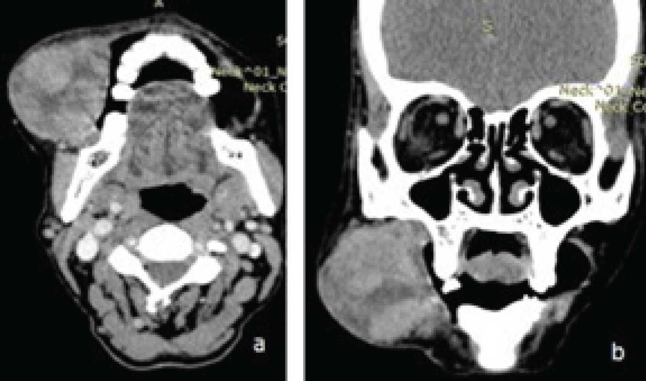
            Figure 3:

            Contrast enhanced computerized tomography (CECT) of face, (a) axial section shows well demarcated lesion on the right cheek originating from the buccal mucosa. There is subtle enhancement with a few regions of necrosis, (b) coronal section of the same lesion highlights non – involvement of the underlying bone and overlying skin.

            Figure 4:

            Magnetic resonance of the same patient, (a) T1W axial image shows well defined hypointense lesion, (b) T2W image shows the lesion as hyperintense with well marginated hypointense capsule, (c) Short tau inversion recovery (STIR) image show the same lesion stands out as hyperintense with the suppression of fat in the background. A few regions of hyperintensities are seen within the pathology representing as necrosis.
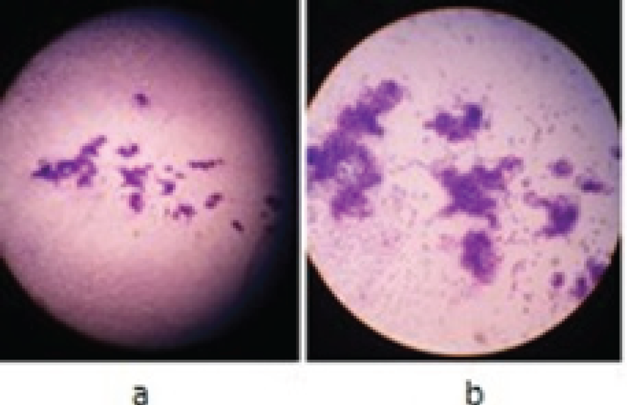
            Figure 5:

            Photomicrograph of the cytology picture, (a) smear shows high cellularity, showing epithelial cells arranged in clusters and singly dispersed at places, Individual cells are showing round to oval nuclei, scant amount of cytoplasm and scant stroma in the background of blood and blood components suggestive of monomorphic adenoma, (b) Magnified image of the same pathology.

            Discussion

            Salivary tumors are slightly uncommon and monomorphic adenomas lists among still less common. Batsakis and Brannon [4] have classified the entity on the histological grounds into the following groups:

            • arising from terminal duct as basal cell adenoma and canalicular adenoma

            • arising from terminal or striated duct as sebaceous adenoma and sebaceous lymphadenoma.

            • arising from striated duct as oncocytoma, papillary cystadenoma and lymphomatosum

            • excretory duct origin tumors as sialadenoma papillefrum or inverted duct papilloma

            Though the exact etiology of these tumors is not known but smoking, vitamin A deficiency and genetic predisposition are considered as the associated factors [5]. The diagnosis of these tumors remains elusive because of their slow growing as well as asymptomatic nature. Ultrasound-guided core needle biopsy is radiation free and safe method for the pathological diagnosis with almost 97% accuracy. This can be performed as outpatient procedure avoiding any hospital stay [6]. The management is for the wider excision of the tumor as there are chances of relapse. The periodic follow-up is necessary in the post-operative period. In malignant pathology, there is role of post-operative radiotherapy with a dose of 60 Gy and of non resectable tumors with 66 Gy [7].

            Conclusion

            The swelling on the cheek can raise various diagnostic dilemmas as the site for epicenter of the origin of tumor becomes difficult. The presenting features of the swelling are delayed because of the painless nature of the entity. The cytopathological features after cross section imaging confirms the diagnosis to unveil the mystery. The proper management can be undertaken if the nature of the tumor and the staging are confirmed. The surgical wide excision is the answer for these tumors with regular follow as these have got tendency for relapse.

            Acknowledgements

            None

            List of Abbreviations

            CECT

            contrast enhanced computerized tomography

            CFI

            Color flow imaging

            FNAC

            fine needle aspiration cytology

            MRI

            magnetic resonance imaging

            US

            Ultrasound

            Conflict of Interests

            None

            Funding

            None

            Consent for publication

            Informed consent was obtained from the patient to present and publish this case.

            Ethical approval

            Ethical approval is not required at our institution for publishing a case report in a medical journal.

            Authors’ contribution

            NB conducted cross-sectional imaging study, SS conducted the clinical examination and wrote the manuscript, MRA designed the manuscript and collected the references, SD provided the images, SN conducted the Histopathological Study.

            All authors contributed in the management of the patient. All of them approved the final version of the manuscript.

            Author details

            Bharat Bhushan Sharma1, Naveen Bhardwaj1, Shweta Sharma2, Mir Rizwan Aziz1, Sakshi Dewan1, Seema Nagger3

            References

            1. Batsakis JG, Brannon RB, Sciubba JJ.. Monomorphic adenomas of major salivary glands: a histologic study of 96 tumours. Clin Otolaryngol Allied Sci. 1981. Vol. 6:129–43

            2. Koyuncu M, Seşen T, Akan H, Ismailoglu AA, Tanyeri Y, Tekat A, et al.. Comparison of computed tomography and magnetic resonance imaging in the diagnosis of parotid tumors. Otolaryngol Head Neck Surg. 2003. Vol. 129:726–32. [10.1016/j.otohns.2003.07.009]

            3. Pinkston JA, Cole P.. Cigarette smoking and Warthin’s tumor. Am J Epidemiol. 1996. Vol. 144:183–7

            4. Terhaard CHJ, Lubsen H, Rasch CRN, Levendag PC, Kaanders HHAM, Tjho-Heslinga RE, et al.. The role of radiotherapy in the treatment of malignant salivary gland tumors. Int J Radiat Oncol Biol Phys. 2005. Vol. 61:103–11. [10.1016/j.ijrobp.2004.03.018]

            5. Wan Y-L, Chan S-C, Chen Y-L, Cheung Y-C, Lui K-W, Wong H-F, et al.. Ultrasonography-guided core-needle biopsy of parotid gland masses. AJNR Am J Neuroradiol. 2004. Vol. 25:1608–12

            6. Yabuuchi H, Fukuya T, Tajima T, Hachitanda Y, Tomita K, Koga M.. Salivary gland tumors: diagnostic valueof gadolinium-enhanced dynamic MR imaging with histopathologic correlation. Radiology. 2003. Vol. 226:345–54. [10.1148/radiol.2262011486]

            7. Yih W-Y, Kratochvil FJ, Stewart JCB.. Intraoral minor salivary gland neoplasms: review of 213 cases. J Oral Maxillofac Surg. 2005. Vol. 63:805–10. [10.1016/j.joms.2005.02.021]

            Summary of the case

            Patient (gender, age) 1Female, 60 year old
            Final Diagnosis 2Buccal Monomorphic Adenoma
            Symptoms 3Swelling over right cheek
            Medications (Generic) 4N/A
            Clinical Procedure 5Fine needle aspiration cytology
            Specialty 6Otorhinolaryngology
            Objective 7To diagnose the swelling over right cheek
            Background 8Elderly female with swelling of right cheek
            Case Report 9Role of cross-sectional imaging in Cheek swelling
            Conclusions 10Sensitivity of Monomorphic adenoma of minor salivary gland
            MeSH Keywords 11Minor salivary glands, monomorphic adenoma, US, CECT, MRI, FNAC

            Author and article information

            Journal
            European Journal of Medical Case Reports
            EJMCR
            Discover STM Publishing Ltd.
            2520-4998
            30 January 2017
            : 1
            : 1
            : 51-55
            Affiliations
            [1 ]Department of Radio-diagnosis, SGT Medical College Budhera (Gurgaon), India.
            [2 ]Department of ENT & Head & Neck Surgery, SGT Medical College Budhera (Gurgaon), India.
            [3 ]Department of Pathology, SGT Medical College Budhera (Gurgaon), India.
            Author notes
            [* ] Correspondence to: Dr. Bharat Bhushan Sharma, Department of Radiodiagnosis, Faculty of Medical and Health Sciences, SGT University Gurgaon (Haryana) India. Email: bbhushan986@ 123456gmail.com
            Article
            ejmcr-1-51
            10.24911/ejmcr/1/13
            21777eae-c61e-402a-953f-c2f68c8c3006
            © Bharat Bhushan Sharma, Naveen Bhardwaj, Shweta Sharma, Mir Rizwan Aziz, Sakshi Dewan, Seema Nagger

            This is an open access article distributed in accordance with the Creative Commons Attribution (CC BY 4.0) license: https://creativecommons.org/licenses/by/4.0/) which permits any use, Share — copy and redistribute the material in any medium or format, Adapt — remix, transform, and build upon the material for any purpose, as long as the authors and the original source are properly cited.

            History
            : 27 November 2016
            : 11 January 2017
            Categories
            CASE REPORT

            FNAC,MRI,CECT,US,monomorphic adenoma,Minor salivary glands

            Comments

            Comment on this article