1,382
views
1
recommends
+1 Recommend
1 collections
    0
    shares

      Acta Materia Medica now indexed by SCOPUS from May 2024. Interested in becoming an AMM published author?

      • Platinum Open Access with no APCs.
      • Fast peer review/Fast publication online after article acceptance.

      Check out the call for papers on our website https://amm-journal.org/index.php/2023/04/26/acta-materia-medica-call-for-papers-2/

      scite_
      0
      0
      0
      0
      Smart Citations
      0
      0
      0
      0
      Citing PublicationsSupportingMentioningContrasting
      View Citations

      See how this article has been cited at scite.ai

      scite shows how a scientific paper has been cited by providing the context of the citation, a classification describing whether it supports, mentions, or contrasts the cited claim, and a label indicating in which section the citation was made.

       
      • Record: found
      • Abstract: found
      • Article: found
      Is Open Access

      Senescence-associated lncRNAs indicate distinct molecular subtypes associated with prognosis and androgen response in patients with prostate cancer

      Published
      research-article
      Bookmark

            Abstract

            Cellular senescence has been considered as a hallmark of aging. In this study, we aimed to establish two novel prognostic subtypes for prostate cancer patients using senescence-related lncRNAs. Nonnegative matrix factorization algorithm was used to identify molecular subtypes. We completed analyses using software R 3.6.3 and its suitable packages. Using SNHG1, MIAT and SNHG3, 430 patients in TCGA database were classified into two subtypes associated with biochemical recurrence (BCR)-free survival and subtype 2 was prone to BCR (HR: 19.62, p < 0.001). The similar results were observed in the GSE46602 and GSE116918. For hallmark gene set enrichment, we found that protein secretion and androgen response were highly enriched in subtype 1 and G2M checkpoint was highly enriched in subtype 2. For tumor heterogeneity and stemness, homologous recombination deficiency and tumor mutation burden were significantly higher in subtype 2 than subtype 1. The top ten genes between subtype 2 and subtype 1 were CUBN, DNAH9, PTCHD4, NOD1, ARFGEF1, HRAS, PYHIN1, ARHGEF2, MYOM1 and ITGB6 with statistical significance. In terms of immune checkpoints, only CD47 was significantly higher in subtype 1 than that in subtype 2. For the overall assessment, no significant difference was detected between two subtypes, while B cells score was significantly higher in subtype 1 than subtype 2. Overall, we found two distinct subtypes closely associated with BCR-free survival and androgen response for prostate cancer. These subtypes might facilitate future research in the field of prostate cancer.

            Main article text

            1. INTRODUCTION

            Bladder, renal and prostate tumors, the most prevalent malignancies in the genitourinary system, are all closely associated with age. Prostate cancer (PCa) continues to be among the top three malignancies in men, in terms of prevalence and death rates (27% and 11%, respectively, in the United States) [1]. Despite major advancements in treatment, PCa continues to pose a serious health risk to men worldwide [24]. Therefore, several therapy options must be considered. One new cancer hallmark included in 2022 is cellular senescence. Senescent cells perform dual roles in a variety of malignant processes [5]. Another recently discovered hallmark of cancer is non-mutational epigenetic reprogramming, in which long non-coding RNAs (lncRNAs) play a key role [5]. Transcripts that are longer than 200 nt and do not encode proteins are known as lncRNAs. These RNAs regulate gene expression at the epigenetic, translational and post-translational levels. Notably, lncRNAs appear to be associated with cellular senescence [68], and increasing evidence indicates critical roles of lncRNA expression in cancers [9, 10]. Studying senescence and lncRNAs in PCa could therefore guide both basic research and clinical treatment.

            LncRNAs are abnormally expressed in a variety of cancers, including PCa. Numerous studies have examined the relationships between lncRNAs and PCa. For instance, prostate cancer antigen 3 (PCA 3), one of the most accurate biomarkers for PCa, functions as a lncRNA and modulates androgen receptor (AR) signal transduction and cell growth [11]. Second chromosomal locus associated with prostate-1 (SChLAP1), a lncRNA that is highly expressed in 25% of PCa, is likewise involved in PCa metastasis and adverse outcomes in patients with PCa [12]. In addition, Singh et al. have recently identified lncRNA H19 as a potential biomarker for neuroendocrine PCa diagnosis and prognostication [13]. They have demonstrated that lncRNAs are useful in both prognosis and treatment, even in patients with the deadly subtype of PCa that is resistant to castration. However, studies investigating that the relationship between lncRNAs and PCa from a perspective of senescence remain lacking.

            In this study, by integrating multiple analytic methods, we constructed and validated two novel prognostic subtypes of patients with PCa by using senescence-associated lncRNAs. The distinct characteristics of these two subtypes regarding prognosis and androgen response may aid in the future PCa research and clinical therapy.

            2. METHODS

            2.1 Data preparation

            We downloaded a list of 279 human genes driving cellular senescence from the CellAge database (http://genomics.senescence.info/cells) [14]. We used the PCa gene matrix and clinical features in The Cancer Genome Atlas (TCGA) database from our pervious study [3]. Biochemical recurrence (BCR)-associated lncRNAs and differentially expressed lncRNAs were examined. Differential expression was defined by a fold-change absolute value >1.5 and an adjusted p value <0.05. The senescence-associated lncRNAs were identified with Pearson analysis, on the basis of a p value <0.5 and absolute value of the coefficient >0.4. A total of 430 samples from the TCGA database were examined, and the log-rank test for BCR-free survival was significant, at p <0.05. Through intersection of differentially expressed, BCR-associated and senescence-associated lncRNAs, we identified lncRNAs that were used to group 430 patients with PCa undergoing radical prostatectomy in TCGA database by using the nonnegative matrix factorization algorithm. We used two additional Gene Expression Omnibus datasets (GSE46602 [15] and GSE116918 [16]) to externally validate the subtypes identified in TCGA database. The prognosis and clinical traits of molecular subtypes were also analyzed.

            2.2 Mutational landscape and functional differences between subtypes

            TCGA (https://portal.gdc.com), a database containing information on PCa, was the source of the downloaded RNA-sequencing profiles, genetic mutations and related clinical data. Using the maftools package in the R programming language, we downloaded and displayed the mutational data. Differences in mutational frequency between subtypes were also assessed. In functional analysis, we performed gene set enrichment analysis with “c2.cp.kegg.v7.4.symbols.gmt” and “h.all.v7.4.symbols.gmt” from the molecular signatures database [17, 18]. The minimal gene set was determined to be 5, whereas the maximum gene set was established to be 5000, on the basis of gene expression and subtypes. Resampling was performed 1000 times. A false discovery rate of 0.10 and a p value of 0.05 were considered to indicate statistical significance.

            2.3 Tumor stemness and heterogeneity analyses

            Tumor stemness indexes included stemness scores based on differentially methylated probes, DNA methylation-based stemness, enhancer element/DNA-methylation-based stemness, epigenetically regulated DNA-methylation-based stemness, epigenetically regulated RNA-expression-based stemness and RNA-expression-based stemness [19]. Tumor heterogeneity included homologous-recombination deficiency (HRD), loss of heterozygosity, neoantigens, tumor ploidy, tumor purity, mutant-allele tumor heterogeneity, tumor mutational burden (TMB) and microsatellite instability [20, 21]. The results of the above indicators were obtained from our previous study [22]. We compared the differences in two subtypes with the Wilcoxon rank-sum test.

            2.4 Tumor microenvironment assessment

            By using the TIMER and ESTIMATE algorithms, we determined the overall tumor microenvironment and immune component assessments [2325]. The Wilcoxon rank-sum test was used to compare the 54 immune-checkpoint differences and tumor microenvironment scores between subtypes. Figure 1 presents the flowchart of our study.

            Figure 1 |

            Study flowchart.

            PCa, prostate cancer; lncRNA, long non-coding RNA; BCR, biochemical recurrence; GSEA, gene set enrichment analysis; NMF, nonnegative matrix factorization.

            2.5 Statistical analysis

            We performed the analysis in R 3.6.3 with the appropriate tools. For data with a non-normal distribution, we used the Wilcoxon rank-sum test. A Kaplan-Meier curve representing the results of the log-rank test was used for survival analysis. The threshold for statistical significance was a two-sided p <0.05. Significance is indicated as follows: not significant (ns), p≥0.05; *, p<0.05; **, p<0.01; *** and p<0.001.

            3. RESULTS

            3.1 Identification of senescence-associated lncRNA subtypes and their applications

            We detected 47 lncRNAs that were differentially expressed between 498 tumor and 52 normal PCa samples in TCGA database ( Figure 2a ). A total of 38 BCR-associated lncRNAs and 73 senescence-associated lncRNAs were detected. Through intersection, we identified 16 differentially expressed lncRNAs associated with BCR and senescence ( Figure 2b ). Subsequently, we used small nucleolar RNA host gene 1 (SNHG1), myocardial infarction associated transcript (MIAT) and small nucleolar RNA host gene 3 (SNHG3) to group the 430 patients in TCGA database, then performed Cox regression analysis including the above 16 lncRNAs ( Figure 2c ). Among the various subtypes ( Figure 2c ), two molecular subtypes were significantly associated with BCR-free survival, and subtype 2 was prone to BCR (HR: 19.62, p <0.001; Figure 3a ). In GSE116918 [16], the three genes were used to divide the 248 patients undergoing radical radiotherapy into two subtypes ( Figure 3b ); subtype 2 had significantly higher risk of BCR than subtype 1 (HR: 27.48, p <0.001; Figure 3c ). Similar results were observed for GSE46602 [15] ( Figure 3d-e ). In addition, the baseline characteristics, e.g., Gleason score and T stage, were balanced between subtypes in both TCGA database ( Table 1 ) and the GSE116918 [16] dataset ( Table 2 ).
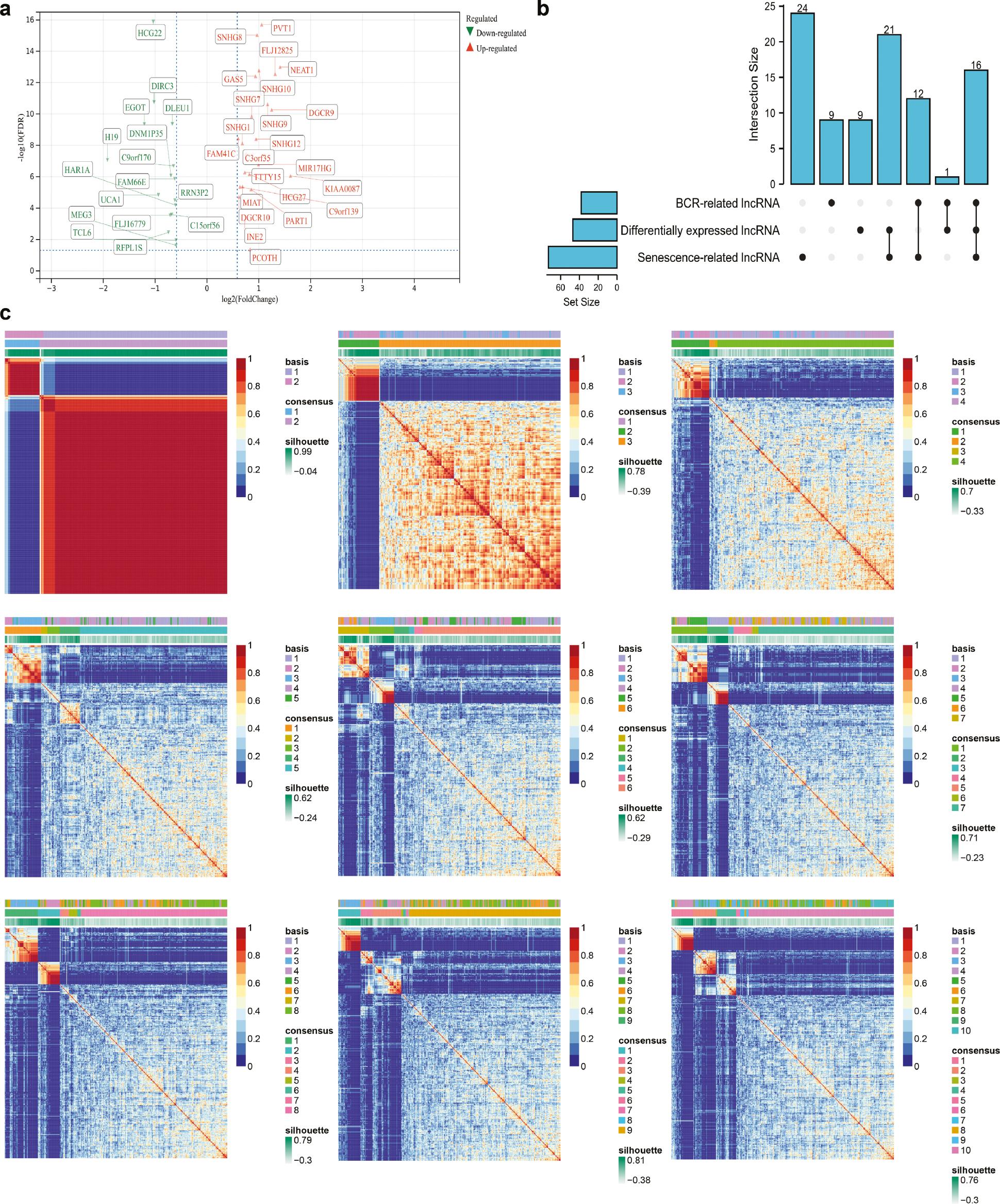
            Figure 2 |

            Identification of TCGA subtypes.

            (a) Volcano plot showing differentially expressed lncRNAs in TCGA database. (b) UpSet plot showing the intersection of senescence-associated, differentially expressed and BCR-associated lncRNAs in the TCGA database. (c) Heatmap plot showing all subtypes in TCGA database, on the basis of the nonnegative matrix factorization algorithm. lncRNA, long non-coding RNA; BCR, biochemical recurrence.

            Figure 3 |

            Prognostic value of subtypes and pathway analysis.

            (a) Kaplan-Meier curve showing the BCR-free survival difference in TCGA database. (b) Heatmap plot showing two distinct subtypes in GSE116918. (c) Kaplan-Meier curve showing the BCR-free survival difference in GSE116918. (d) Heatmap plot showing two distinct subtypes in the GSE46602. (e) Kaplan-Meier curve showing the BCR-free survival difference in GSE46602. (f) Hallmark gene set analysis of TCGA subtypes. (g) KEGG pathway differences in TCGA subtypes. (h) Forest plot showing results of tumor heterogeneity and stemness between TCGA subtypes. (i) Waterfall plot showing the top ten differentially mutated genes between TCGA subtypes. BCR, biochemical recurrence.

            Table 1 |

            Differences in clinical characteristics between two prostate cancer subtypes in TCGA.

            CharacteristicSubtype 1Subtype 2p Value
            Samples35575
            Age, median (IQR)61 (56, 66)62 (57, 66)0.495
            Gleason score, n (%)0.147
             632 (7.4%)7 (1.6%)
             7177 (41.2%)29 (6.7%)
             850 (11.6%)9 (2.1%)
             996 (22.3%)30 (7%)
            T stage, n (%)1.000
             T2128 (30.2%)27 (6.4%)
             T3215 (50.7%)46 (10.8%)
             T47 (1.7%)1 (0.2%)
            Race, n (%)0.317
             Asian9 (2.2%)2 (0.5%)
             Black or African American45 (10.8%)5 (1.2%)
             White288 (69.2%)67 (16.1%)
            N stage, n (%)0.901
             N0253 (67.5%)53 (14.1%)
             N156 (14.9%)13 (3.5%)
            Residual tumor, n (%)0.272
             No230 (54.9%)43 (10.3%)
             Yes116 (27.7%)30 (7.2%)

            IQR, interquartile range.

            Table 2 |

            Differences in clinical characteristics between prostate cancer subtypes in GSE116918.

            CharacteristicSubtype 1Subtype 2p Value
            Sample22622
            Age, median (IQR)68 (63.25, 72)69.5 (62.5, 73)0.534
            T stage, n (%)0.095
             T148 (21.5%)3 (1.3%)
             T269 (30.9%)7 (3.1%)
             T383 (37.2%)9 (4%)
             T42 (0.9%)2 (0.9%)
            Gleason score, n (%)0.075
             642 (16.9%)0 (0%)
             789 (35.9%)10 (4%)
             845 (18.1%)7 (2.8%)
             950 (20.2%)5 (2%)
            BCR, n (%)<0.001
             No182 (73.4%)10 (4%)
             Yes44 (17.7%)12 (4.8%)
            Metastasis, n (%)0.001
             No211 (85.1%)15 (6%)
             Yes15 (6%)7 (2.8%)

            IQR, interquartile range.

            3.2 Functional enrichment, mutated genes, tumor heterogeneity and stemness

            In hallmark gene set enrichment, protein secretion and AR were highly enriched in subtype 1, and the G2M checkpoint was highly enriched in subtype 2 ( Figure 3f ). In pathway analysis, spliceosomes, base excision repair and the cell cycle were highly enriched in subtype 2 ( Figure 3g ). Regarding tumor heterogeneity and stemness, HRD and TMB were significantly higher in subtype 2 than subtype 1 ( Figure 3h ). The top ten significantly differentially expressed genes between subtype 2 and subtype 1 were CUBN, DNAH9, PTCHD4, NOD1, ARFGEF1, HRAS, PYHIN1, ARHGEF2, MYOM1 and ITGB6 ( Figure 3i ).

            3.3 Tumor immune microenvironment and immune checkpoints

            Among immune checkpoints, the expression levels of CTLA4, ICOS, TNFRSF18, TNFRSF4, TNFRSF25, LAG3, TNFRSF8, CD80, ADORA2A and CD276 were significantly higher in subtype 2 than in subtype 1 ( Figure 4a ). Only CD47 was significantly higher in subtype 1 than subtype 2 ( Figure 4a ). In the overall assessment, no significant difference was detected between subtypes, whereas the B cell scores were significantly higher in subtype 1 than subtype 2 ( Figure 4b ).

            Figure 4 |

            TME and immune-checkpoint analysis.

            (a) Forest plot showing significant differences in two TCGA subtypes in terms of checkpoints. (b) Forest plot showing significant differences in two TCGA subtypes for TME assessment. TME, tumor immune microenvironment; CI, confidence interval.

            4. DISCUSSION

            After thorough validation, we identified two distinct prognosis-associated subgroups of patients with PCa. Subtype 2, with a poorer prognosis, was distinguished from subtype 1 by the lncRNAs SNHG1, MIAT and SNHG3. Importantly, according to our hallmark gene set enrichment analysis, this typing system aids in identifying patients who are not susceptible to androgens. Additionally, the main differences between subtypes and their probable causes were determined through investigation of tumor heterogeneity, the mutational landscape and PCa immunity.

            SNHG1, MIAT and SNHG3 were lncRNAs differentially expressed between cancerous tissues and adjacent normal tissues. SNHG1 is a novel lncRNA whose high expression and carcinogenic characteristics have been confirmed in various cancers [2629]. In PCa, SNHG1 mediates malignant transformation by promoting tumor proliferation, invasion and epithelial-mesenchymal transition, primarily through a competing endogenous RNA mechanism [2931]. Additionally, studies on SNHG3 and PCa have shown similar results to studies on SNHG1. Interactions between SNHG3 and microRNA epigenetically regulate PCa progression [3234]. Although the role of MIAT in PCa remains to be clarified, Crea et al. have suggested that MIAT also promotes neuroendocrine PCa initiation and progression [35]. Nonetheless, our experimental data and validation suggested that patients in subtype 2 were prone to BCR, on the basis of typing according to those three lncRNAs.

            Because lncRNAs function primarily as epigenetic modulators, we conducted hallmark gene set enrichment analysis. We observed clear enrichment in genes associated with AR in subtype 1, in which patients had better prognosis. Androgen dependence is a typical characteristic of primary PCa [36]. After ARs receive signals from androgens and associated products, they initiate the activation and transduction of a series of signaling pathways via a complex intracellular signaling network. Mechanically, AR is a transcription factor involved in the cell cycle, prostate development and other key activities, and it has been found to promote PCa growth [37]. Therefore, androgen-deprivation therapy was the traditional treatment for PCa [38]. However, patients with PCa who undergo androgen-deprivation therapy subsequently experience recurrence and therapy resistance, and develop castration-resistant PCa after 18–36 months [39, 40]. Androgen-independent abnormal reactivation of AR signals results in such cancer progression [41]. Although novel second-generation AR antagonists, such as enzalutamide and abiraterone, may work temporarily, patients still encounter inevitable drug resistance, disease progression and even lethal outcomes [38]. Hence, androgen sensitivity and response are critical in the prognosis of patients with PCa. Accordingly, enriched AR genes may be considered the key factors associated with the better prognosis in subtype 1. Moreover, a recent study has shown indicated that molecular modulation and interactions between other proteins and DNAs are involved in the resistance to enzalutamide and abiraterone [42]. Accordingly, we hypothesized that altered expression of the three lncRNAs in subtype 2 might also account for the altered AR characteristics and consequently the poor prognosis, from an AR-epigenetic modulation perspective. In contrast, G2M-checkpoint-associated genes were enriched in subtype 2. Tumors cannot grow without constant and infinite mitosis, and the G2M checkpoint determines whether cells enter the M stage of the cell cycle. Several lncRNA have been confirmed to interfere with the normal cell cycle by targeting the G2M checkpoint [4345]. Subsequently, our hypothesis that the expression of the lncRNAs SNHG1, MIAT and SNHG3 promotes malignant proliferation in PCa may be a reasonable explanation for the short BCR-free survival among patients with subtype 2 PCa. Our pathway analysis provided evidence supporting that possibility: pathways associated with cell proliferation and cell death, including spliceosomes, base excision repair and the cell cycle, were highly enriched in subtype 2. Together, the three lncRNAs identified herein may regulate AR activity and the cell cycle in PCa cells, thus providing a basis for our subtyping analysis.

            We also identified mutated genes in the two subtypes: CUBN, DNAH9 and PTCHD4 were the top three mutation genes. CUBN, a co-transporter located primarily in absorptive epithelia, promotes the uptake of specific ligands, such as hemoglobin, lipoprotein and iron [46]. The expression of CUBN is associated with the initiation and progression of renal clear cell carcinoma, colorectal cancer and breast cancer [4749]; moreover, low CUBN expression is significantly associated with poor prognosis [49]. The loss of DNAH9 often occurs in tumors, and Donner et al. have revealed that DNAH9 may possess antitumor effects [50]. Furthermore, epigenetic regulation of PTCHD4 splicing decreases tumor growth, thus indicating a potential role of PTCHD4 in PCa [51]. Targeting these top mutated genes might improve the prognosis of subtype 2 patients.

            Because tumor immunity has non-negligible effects on patient prognosis, we analyzed several immunity-associated indicators to better interpret our typing mechanisms. In the tumor immune microenvironment (TME), immune silencing and immunosuppressive status have long been considered characteristics of PCa immunity [52]. In our TME score analysis, we observed that infiltrating immune and immune-associated cells in subtype 2 did not substantially differ from those in subtype 1 (except for B cells), and the difference in immune infiltrating status between subtypes was not sufficient to alter patient prognosis. However, our subsequent findings may advance current viewpoints regarding PCa immunotherapy. The TMB and HRD were significantly higher in subtype 2 than subtype 1. TMB indicates the total number of mutations and the degree of tumor heterogeneity [20, 21]. On the basis of the hypothesis that mutant proteins would increase the number of antigenic peptides and generate new immunogenic antigens [53, 54], TMB may be applied to predict therapeutic responses to immune-checkpoint blockade and identify patients who might benefit most from treatment. Importantly, increasing evidence suggests that cellular heterogeneity does not simply result from transcriptional processes but instead has an epigenetic basis, in which histone modifications, nucleosome positioning and chromatin accessibility have been demonstrated to be involved, and lncRNAs may also play a role [55]. In addition, HRD is a key therapy response indicator that results in specific and stable genomic changes in PCa [56, 57]. Changes in the two indicators of tumor heterogeneity may suggest that our typing system could be applied to identify specific patient populations that might benefit most from immunotherapy. In addition, most significantly differentially expressed immune checkpoints in our analysis had higher expression in subtype 2 than subtype 1, thereby suggesting a relatively more active immune TME with more potential therapy targets in subtype 2.

            In summary, our study established two novel prognostic subtypes that were closely associated with BCR-free survival in patients with PCa, and suggested that the AR may play a critical role. Our typing system additionally indicated immune status. Therefore, our study may guide the clinical management of patients with PCa. However, further studies and experiments will be necessary to reveal the detailed mechanisms.

            5. CONCLUSION

            In conclusion, we identified two distinct subtypes closely associated with BCR-free survival and AR for PCa, which might aid in future research in the field of PCa.

            CONFLICTS OF INTERESTS

            The authors declare that there are no conflicts of interest.

            REFERENCES

            1. Siegel RL, Miller KD, Fuchs HE, Jemal A. Cancer Statistics, 2022. CA: A Cancer Journal for Clinicians. 2022. Vol. 72:7–33

            2. Feng D, Xiong Q, Wei Q, Yang L. Cellular Landscape of Tumour Microenvironment in Prostate Cancer. Immunology. 2023. Vol. 168:199–202

            3. Feng D, Shi X, Zhang F, Xiong Q, Wei Q, Yang L. Energy Metabolism-Related Gene Prognostic Index Predicts Biochemical Recurrence for Patients with Prostate Cancer Undergoing Radical Prostatectomy. Frontiers in Immunology. 2022. Vol. 13:839362

            4. Zi H, He SH, Leng XY, Xu XF, Huang Q, Weng H, et al.. Global, Regional, and National Burden of Kidney, Bladder, and Prostate Cancers and their Attributable Risk Factors, 1990-2019. Military Medical Research. 2021. Vol. 8:60

            5. Hanahan D. Hallmarks of Cancer: New Dimensions. Cancer Discovery. 2022. Vol. 12:31–46

            6. Haemmig S, Yang D, Sun X, Das D, Ghaffari S, Molinaro R, et al.. Long Noncoding RNA SNHG12 Integrates a DNA-PK-Mediated DNA Damage Response and Vascular Senescence. Science Translational Medicine. 2020. Vol. 12:eaaw1868

            7. Nojima T, Tellier M, Foxwell J, Ribeiro de Almeida C, Tan-Wong SM, Dhir S, et al.. Deregulated Expression of Mammalian lncRNA through Loss of SPT6 Induces R-Loop Formation, Replication Stress, and Cellular Senescence. Molecular Cell. 2018. Vol. 72:970–984.e7

            8. Yang Z, Jiang S, Shang J, Jiang Y, Dai Y, Xu B, et al.. LncRNA: Shedding Light on Mechanisms and Opportunities in Fibrosis and Aging. Ageing Research Reviews. 2019. Vol. 52:17–31

            9. Bhan A, Soleimani M, Mandal SS. Long Noncoding RNA and Cancer: A New Paradigm. Cancer Research. 2017. Vol. 77:3965–3981

            10. Tan YT, Lin JF, Li T, Li JJ, Xu RH, Ju HQ. LncRNA-Mediated Posttranslational Modifications and Reprogramming of Energy Metabolism in Cancer. Cancer Communications (London, England). 2021. Vol. 41:109–120

            11. Salameh A, Lee AK, Cardó-Vila M, Nunes DN, Efstathiou E, Staquicini FI, et al.. PRUNE2 is a Human Prostate Cancer Suppressor Regulated by the Intronic Long Noncoding RNA PCA3. Proceedings of the National Academy of Sciences of the United States of America. 2015. Vol. 112:8403–8408

            12. Prensner JR, Iyer MK, Sahu A, Asangani IA, Cao Q, Patel L, et al.. The Long Noncoding RNA SChLAP1 Promotes Aggressive Prostate Cancer and Antagonizes the SWI/SNF Complex. Nature Genetics. 2013. Vol. 45:1392–1398

            13. Singh N, Ramnarine VR, Song JH, Pandey R, Padi SKR, Nouri M, et al.. The Long Noncoding RNA H19 Regulates Tumor Plasticity in Neuroendocrine Prostate Cancer. Nature Communications. 2021. Vol. 12:7349

            14. Avelar RA, Ortega JG, Tacutu R, Tyler EJ, Bennett D, Binetti P, et al.. A Multidimensional Systems Biology Analysis of Cellular Senescence in Aging and Disease. Genome Biology. 2020. Vol. 21:91

            15. Mortensen MM, Høyer S, Lynnerup AS, Ørntoft TF, Sørensen KD, Borre M, et al.. Expression Profiling of Prostate Cancer Tissue Delineates Genes Associated with Recurrence after Prostatectomy. Scientific Reports. 2015. Vol. 5:16018

            16. Jain S, Lyons CA, Walker SM, McQuaid S, Hynes SO, Mitchell DM, et al.. Validation of a Metastatic Assay using Biopsies to Improve Risk Stratification in Patients with Prostate Cancer Treated with Radical Radiation Therapy. Annals of Oncology. 2018. Vol. 29:215–222

            17. Liberzon A, Subramanian A, Pinchback R, Thorvaldsdóttir H, Tamayo P, Mesirov JP. Molecular Signatures Database (MSigDB) 3.0. Bioinformatics. 2011. Vol. 27:1739–1740

            18. Subramanian A, Tamayo P, Mootha VK, Mukherjee S, Ebert BL, Gillette MA, et al.. Gene Set Enrichment Analysis: A Knowledge-based Approach for Interpreting Genome-Wide Expression Profiles. Proceedings of the National Academy of Sciences of the United States of America. 2005. Vol. 102:15545–15550

            19. Malta TM, Sokolov A, Gentles AJ, Burzykowski T, Poisson L, Weinstein JN, et al.. Machine Learning Identifies Stemness Features Associated with Oncogenic Dedifferentiation. Cell. 2018. Vol. 173:338–354.e15

            20. Bonneville R, Krook MA, Kautto EA, Miya J, Wing MR, Chen HZ, et al.. Landscape of Microsatellite Instability Across 39 Cancer Types. JCO Precision Oncology. 2017. Vol. 2017:PO.17.00073

            21. Thorsson V, Gibbs DL, Brown SD, Wolf D, Bortone DS, Ou Yang TH, et al.. The Immune Landscape of Cancer. Immunity. 2018. Vol. 48:812–830.e14

            22. Feng D, Shi X, Zhu W, Zhang F, Li D, Han P, et al.. A Pan-Cancer Analysis of the Oncogenic Role of Leucine Zipper Protein 2 in Human Cancer. Experimental Hematology and Oncology. 2022. Vol. 11:55

            23. Li B, Severson E, Pignon JC, Zhao H, Li T, Novak J, et al.. Comprehensive Analyses of Tumor Immunity: Implications for Cancer Immunotherapy. Genome Biology. 2016. Vol. 17:174

            24. Yoshihara K, Shahmoradgoli M, Martinez E, Vegesna R, Kim H, Torres-Garcia W, et al.. Inferring Tumour Purity and Stromal and Immune Cell Admixture from Expression Data. Nature Communications. 2013. Vol. 4:2612

            25. Zeng D, Ye Z, Shen R, Yu G, Wu J, Xiong Y, et al.. IOBR: Multi-Omics Immuno-Oncology Biological Research to Decode Tumor Microenvironment and Signatures. Frontiers in Immunology. 2021. Vol. 12:687975

            26. Li W, Dong X, He C, Tan G, Li Z, Zhai B, et al.. LncRNA SNHG1 Contributes to Sorafenib Resistance by Activating the Akt Pathway and is Positively Regulated by miR-21 in Hepatocellular Carcinoma Cells. Journal of Experimental and Clinical Cancer Research. 2019. Vol. 38:183

            27. Thin KZ, Tu JC, Raveendran S. Long Non-Coding SNHG1 in Cancer. Clinica Chimica Acta. 2019. Vol. 494:38–47

            28. Xu M, Chen X, Lin K, Zeng K, Liu X, Pan B, et al.. The Long Noncoding RNA SNHG1 Regulates Colorectal Cancer Cell Growth through Interactions with EZH2 and miR-154-5p. Molecular Cancer. 2018. Vol. 17:141

            29. Meng XF, Liu AD, Li SL. SNHG1 Promotes Proliferation, Invasion and EMT of Prostate Cancer Cells through miR-195-5p. European Review for Medical and Pharmacological Sciences. 2020. Vol. 24:9880–9888

            30. Tan X, Chen WB, Lv DJ, Yang TW, Wu KH, Zou LB, et al.. LncRNA SNHG1 and RNA Binding Protein hnRNPL form a Complex and Coregulate CDH1 to Boost the Growth and Metastasis of Prostate Cancer. Cell Death and Disease. 2021. Vol. 12:138

            31. Li J, Zhang Z, Xiong L, Guo C, Jiang T, Zeng L, et al.. SNHG1 lncRNA Negatively Regulates miR-199a-3p to Enhance CDK7 Expression and Promote Cell Proliferation in Prostate Cancer. Biochemical and Biophysical Research Communications. 2017. Vol. 487:146–152

            32. Hu M, Ren M, Zhao Z, Cui X, Shi M, Yang Y, et al.. Long Non-Coding RNA SNHG3 Promotes Prostate Cancer Progression by Sponging microRNA-1827. Oncology Letters. 2022. Vol. 24:281

            33. Li T, Xing Y, Yang F, Sun Y, Zhang S, Wang Q, et al.. LncRNA SNHG3 Sponges miR-577 to Up-Regulate SMURF1 Expression in Prostate Cancer. Cancer Medicine. 2020. Vol. 9:3852–3862

            34. Yu L, Ren Y. Long Noncoding RNA Small Nucleolar RNA Host Gene 3 Mediates Prostate Cancer Migration, Invasion, and Epithelial-Mesenchymal Transition by Sponging miR-487a-3p to Regulate TRIM25. Cancer Biotherapy and Radiopharmaceuticals. 2022. Vol. 37:451–465

            35. Crea F, Venalainen E, Ci X, Cheng H, Pikor L, Parolia A, et al.. The Role of Epigenetics and Long Noncoding RNA MIAT in Neuroendocrine Prostate Cancer. Epigenomics. 2016. Vol. 8:721–731

            36. Huggins C, Hodges CV. Studies on Prostatic Cancer. I. The Effect of Castration, of Estrogen and Androgen Injection on Serum Phosphatases in Metastatic Carcinoma of the Prostate. CA: A Cancer Journal for Clinicians. 1972. Vol. 22:232–240

            37. Snow O, Lallous N, Singh K, Lack N, Rennie P, Cherkasov A, et al.. Androgen Receptor Plasticity and its Implications for Prostate Cancer Therapy. Cancer Treatment Reviews. 2019. Vol. 81:101871

            38. Teo MY, Rathkopf DE, Kantoff P. Treatment of Advanced Prostate Cancer. Annual Review of Medicine. 2019. Vol. 70:479–499

            39. Yap TA, Smith AD, Ferraldeschi R, Al-Lazikani B, Workman P, de Bono JS. Drug Discovery in Advanced Prostate Cancer: Translating Biology into Therapy. Nature Reviews, Drug Discovery. 2016. Vol. 15:699–718

            40. Sumanasuriya S, De Bono J. Treatment of Advanced Prostate Cancer-A Review of Current Therapies and Future Promise. Cold Spring Harbor Perspectives in Medicine. 2018. Vol. 8:a030635

            41. Shafi AA, Yen AE, Weigel NL. Androgen Receptors in Hormone-Dependent and Castration-Resistant Prostate Cancer. Pharmacology and Therapeutics. 2013. Vol. 140:223–238

            42. Sawant M, Mahajan K, Renganathan A, Weimholt C, Luo J, Kukshal V, et al.. Chronologically Modified Androgen Receptor in Recurrent Castration-Resistant Prostate Cancer and its Therapeutic Targeting. Science Translational Medicine. 2022. Vol. 14:eabg4132

            43. Fu Y, Wei X, Han Q, Le J, Ma Y, Lin X, et al.. Identification and Characterization of a 25-lncRNA Prognostic Signature for Early Recurrence in Hepatocellular Carcinoma. BMC Cancer. 2021. Vol. 21:1165

            44. Pan X, Wu J, Jiang C, Yu Q, Yan B. Synergistic Effects of Carbon Nanoparticle-Cr-Pb in PM(2.5) Cause Cell Cycle Arrest via Upregulating a Novel lncRNA NONHSAT074301.2 in Human Bronchial Epithelial Cells. Journal of Hazardous Materials. 2021. Vol. 411:125070

            45. Xia W, Zeng C, Zheng Z, Huang C, Zhou Y, Bai L. Development and Validation of a Novel Mitochondrion and Ferroptosis-Related Long Non-Coding RNA Prognostic Signature in Hepatocellular Carcinoma. Frontiers in Cell and Developmental Biology. 2022. Vol. 10:844759

            46. Shaik AP, Alsaeed AH, Kiranmayee S, Bammidi V, Sultana A. Phylogenetic Analysis of Cubilin (CUBN) Gene. Bioinformation. 2013. Vol. 9:29–36

            47. Wu Y, Xu Y. Bioinformatics for the Prognostic Value and Function of Cubilin (CUBN) in Colorectal Cancer. Medical Science Monitor. 2020. Vol. 26:e922447

            48. Wang H, Zhao L, Liu H, Luo S, Akinyemiju T, Hwang S, et al.. Variants in SNAI1, AMDHD1 and CUBN in Vitamin D Pathway Genes are Associated with Breast Cancer Risk: A Large-Scale Analysis of 14 GWASs in the DRIVE Study. American Journal of Cancer Research. 2020. Vol. 10:2160–2173

            49. Gremel G, Djureinovic D, Niinivirta M, Laird A, Ljungqvist O, Johannesson H, et al.. A Systematic Search Strategy Identifies Cubilin as Independent Prognostic Marker for Renal Cell Carcinoma. BMC Cancer. 2017. Vol. 17:9

            50. Donner I, Katainen R, Tanskanen T, Kaasinen E, Aavikko M, Ovaska K, et al.. Candidate Susceptibility Variants for Esophageal Squamous Cell Carcinoma. Genes, Chromosomes and Cancer. 2017. Vol. 56:453–459

            51. Huang MH, Chou YW, Li MH, Shih TE, Lin SZ, Chuang HM, et al.. Epigenetic Targeting DNMT1 of Pancreatic Ductal Adenocarcinoma using Interstitial Control Release Biodegrading Polymer Reduced Tumor Growth through Hedgehog Pathway Inhibition. Pharmacological Research. 2019. Vol. 139:50–61

            52. Majidpoor J, Mortezaee K. The Efficacy of PD-1/PD-L1 Blockade in Cold Cancers and Future Perspectives. Clinical Immunology. 2021. Vol. 226:108707

            53. Chan TA, Yarchoan M, Jaffee E, Swanton C, Quezada SA, Stenzinger A, et al.. Development of Tumor Mutation Burden as an Immunotherapy Biomarker: Utility for the Oncology Clinic. Annals of Oncology. 2019. Vol. 30:44–56

            54. Gubin MM, Artyomov MN, Mardis ER, Schreiber RD. Tumor Neoantigens: Building a Framework for Personalized Cancer Immunotherapy. The Journal of Clinical Investigation. 2015. Vol. 125:3413–3421

            55. Carter B, Zhao K. The Epigenetic Basis of Cellular Heterogeneity. Nature Reviews, Genetics. 2021. Vol. 22:235–250

            56. Nguyen L, Martens JWM, Van Hoeck A, Cuppen E. Pan-Cancer Landscape of Homologous Recombination Deficiency. Nature Communications. 2020. Vol. 11:5584

            57. Mateo J, Carreira S, Sandhu S, Miranda S, Mossop H, Perez-Lopez R, et al.. DNA-Repair Defects and Olaparib in Metastatic Prostate Cancer. The New England Journal of Medicine. 2015. Vol. 373:1697–1708

            Graphical abstract

            Highlights
            • We proposed senescence-associated lncRNA-mediated molecular subtypes for prostate cancer.

            • The two subtypes of prostate cancer were associated with biochemical recurrence-free survival and androgen response.

            • The key lncRNAs identified in this study might serve as prognostic biomarkers for prostate cancer.

            In brief

            We found two distinct subtypes closely associated with biochemical recurrence-free survival and androgen response for prostate cancer, which might be important for furture research in the field of prostate cancer.

            Author and article information

            Journal
            amm
            Acta Materia Medica
            Compuscript (Ireland )
            2737-7946
            10 August 2023
            : 2
            : 3
            : 299-309
            Affiliations
            [a ]Department of Urology, Institute of Urology, West China Hospital, Sichuan University, Chengdu 610041, P.R. China
            [b ]Department of Rehabilitation, The Affiliated Hospital of Southwest Medical University, Luzhou 646000, P.R. China
            Author notes
            *Correspondence: fdcfenix@ 123456stu.scu.edu.cn , dechaofeng66@ 123456swmu.edu.cn , Tel.: +86-28-85422444, Fax: +86-28-85422451 (D. Feng); chizhang_swmu@ 123456126.com (C. Zhang)

            1These authors contributed equally to this work.

            Article
            10.15212/AMM-2023-0025
            8e55fa0d-917b-46a0-881e-65c4838ccecf
            Copyright © 2023 The Authors.

            Creative Commons Attribution 4.0 International License

            History
            : 10 July 2023
            : 20 July 2023
            : 30 July 2023
            Page count
            Figures: 4, Tables: 2, References: 57, Pages: 11
            Funding
            Funded by: Luzhou City Science and Technology Bureau
            Award ID: 2021LZXNYDJ10
            Funded by: Luzhou City Science and Technology Bureau
            Award ID: 2020LZXNYDJ14
            Funded by: Cooperation Project between the Second People’s Hospital of Deyang and Southwest Medical University
            Award ID: 2022DYEXNYD002
            We appreciate Chengdu Basebiotech Co, Ltd for assistance in figure generation and data processing. This program was supported by the Luzhou City Science and Technology Bureau (grant number 2021LZXNYDJ10 and 2020LZXNYDJ14), Cooperation Project between the Second People’s Hospital of Deyang and Southwest Medical University (2022DYEXNYD002). The funders had no role in the study design, data collection or analysis, preparation of the manuscript or the decision to publish.
            Categories
            Research Article

            Toxicology,Pathology,Biochemistry,Clinical chemistry,Pharmaceutical chemistry,Pharmacology & Pharmaceutical medicine
            biochemical recurrence,androgen response,molecular subtypes,nonnegative matrix factorization,cellular senescence,prostate cancer

            Comments

            Comment on this article